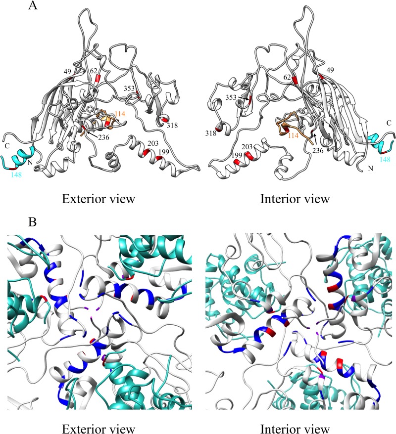
FIG 6.
Locations of the second-site suppressors. (A) The viral coat protein is depicted gray, the internal scaffolding protein in orange, and the D4 external scaffolding protein unit in cyan. There are four D protein subunits per asymmetric unit. Only a portion of the scaffolding proteins are depicted. The residues in which the suppressors reside are depicted in red. The color of the numerical labels indicates the affected protein and the suppressor's location in the primary structure. The side chains of residues F67, Y134, and F135 in F protein and F120 in B protein are included to identify the core of the binding cleft. (B) The 3-fold axis of symmetry. The blue, red, and purple colors identify the locations of the suppressors. Red indicates suppressors identified in this study. Blue indicates suppressors of defective external scaffolding protein function isolated in previous studies. Purple indicates those suppressors identified in this study that were identical and independently isolated in previous studies.

