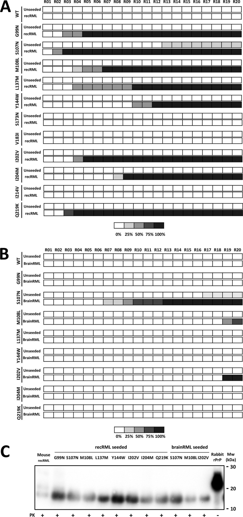
FIG 3.

Three of 11 recombinant rabbit PrPs with mouse PrP single-residue substitutions were misfolded by recRML and brainRML murine prions in vitro. (A) Graphical representation of the emergence and relative amount of protein misfolding (PrPres) for each round of recPMCA (denoted R01 to R20) as evaluated by Western blotting and SDS-PAGE. The percentages of tubes showing PK-resistant misfolded rec-PrP are indicated in grayscale according to the legend. Different mutated rabbit recombinant proteins were subjected to serial rounds of recPMCA after seeding with recRML (misfolded recombinant mouse PrP [recRML-High]). Note the spontaneous emergence of misfolding for the unseeded S107N variant. WT, wild-type rabbit rec-PrPs. (B) Selected mutated rabbit recombinant proteins were subjected to serial rounds of recPMCA after seeding with brainRML (brain-derived RML). Every experiment contained 4 tubes (intraexperimental replicates). The percentage of positive tubes (tubes showing a protease K-resistant signal after digestion with 80 μg/ml of PK) after each round of recPMCA is noted in grayscale as shown in the legend. (C) Western blot representing the recRML (recombinant origin)- or brainRML (brain origin)-seeded and misfolded mutated rabbit rec-PrPs after PK digestion. Samples were digested with 85 μg/ml of PK. Similar bands of approximately 17 kDa, corresponding to the PK-resistant 90-230 fragment of the prion protein, are shown. Membranes were developed with monoclonal antibody SAF84 (1:400). Rabbit rPrP, undigested recombinant rabbit rec-PrP.
