Figure 5.

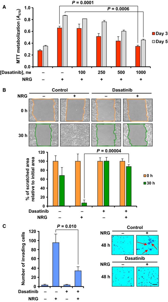
Dasatinib inhibits NRG‐induced proliferation, migration, and invasion. (A) Bar graph representing the dose–response effect of dasatinib on NRG‐induced proliferation in MCF7 cells. Cell proliferation was determined by MTT metabolization 3 and 5 days after NRG stimulation. Four different concentrations (ranging from 100 to 1000 nm) were used. (B) Representative images from the wound healing assay showing the effect of dasatinib on NRG‐induced migration of MCF7 cells at 0 and 30 h after NRG stimulation. The colored lines delimit the area of the wounded region. The bar graph at the bottom represents the quantitation of the wounded area with respect to the initial area in each condition (0 h). (C) Bar graph showing the number of MCF7 invading cells 48 h after NRG stimulation and the effect of pretreatment with dasatinib. Cells that were able to pass through the Matrigel layer were fixed, stained with crystal violet, and counted. Representative images of the experiment are shown on the right. The arrows point to cells stained with crystal violet. Scale bar = 100 μm. Data information: Results are presented as the mean ± SD of triplicate of an experiment that was repeated three times. Comparisons of means between two independent groups were made using a two‐sided Student's t‐test.
