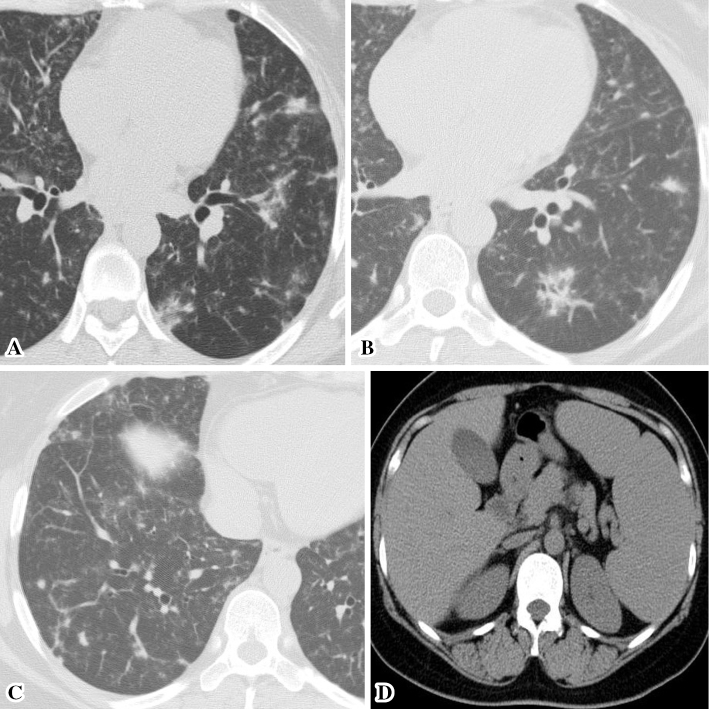
Figure 1.
Radiologic findings. Chest and abdominal computed tomography revealed multiple areas of patchy consolidation and ill-defined nodules along the lymphatic channels, predominantly in the lower lung lobes, thickening of the bronchovascular bundle and interlobular septa (A-C), and splenomegaly (D).

