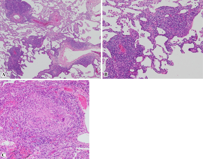
Figure 2.
Pathologic findings. A, B: The pathologic findings of the VATS specimen show infiltration of polyclonal lymphocytes and lymphoid hyperplasia along the bronchi, bronchioles, and vessels, but no lymphoepithelial lesions [A: Hematoxylin and Eosin (H&E) staining ×40, B: H&E staining ×100]. C: A small number of non-caseous epithelioid cell granulomas are observed (H&E staining ×400).

