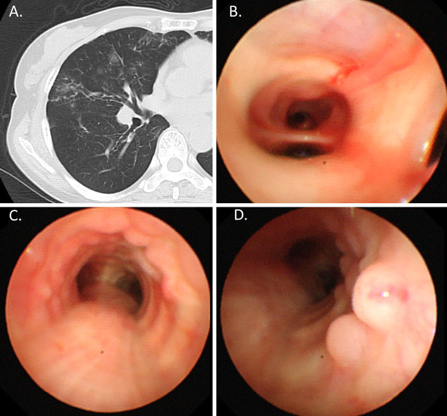
Figure 3.
A: Lung window. B, C, and D: Bronchoscopic findings. Chest CT showing small pulmonary nodules and bronchiectasis in the middle lobe (A). Bronchoalveolar lavage from the middle lobe (B). Bronchoscopic findings revealing multiple small nodular mucosal lesions with a cobblestone appearance along the trachea (C and D).

