-
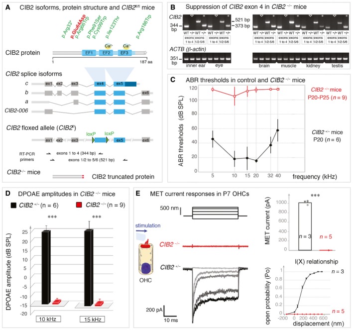
A
Domain structure of the CIB2 protein, indicating the positions of the CIB2 mutations in USH1J (red) and DFNB48 (green) patients. The CIB2‐floxed mice, CIB2
fl/fl, were engineered by adding LoxP sites on either side of exon 4, which is common to all four known CIB2 transcripts.
-
B
RT–PCR analysis confirming the loss of CIB2 exon 4‐containing transcripts in the inner ear, eye, brain, muscle, kidney and testis of CIB2
−/− mice. β‐Actin was used as an endogenous control.
-
C, D
ABR thresholds (C) and DPOAE amplitudes (D) in CIB2
+/− (dark, n = 6) and CIB2
−/− (red, n = 9) P20‐P25 mice. ABR thresholds in CIB2
−/− mice exceeded 100 dB SPL (mean ± SD), indicating profound deafness. (D) DPOAE amplitudes were absent in CIB2
−/− P20 mice at 10 and 15 kHz (red) (Mann–Whitney, ***P = 0.002 for both 10 and 15 kHz).
-
E
MET responses in OHCs from CIB2
+/− and CIB2
−/− P7 mice. The left panels show the mechanical stimulation protocol, with examples of MET currents for each genotype. In the right panels, the MET current values and mean amplitude‐displacement relationships (I(X)) (mean ± SD) in CIB2
+/− (black, n = 3 cells) and CIB2
−/− (red, n = 5) mice highlight the absence of a MET response in CIB2
−/− OHCs (Welch's unpaired t‐test, ****P = 0.0007).