
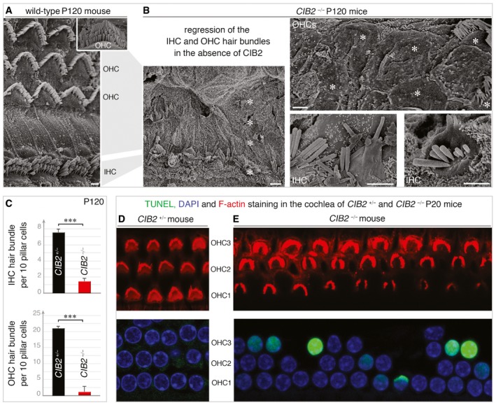
Figure 5. Loss of cochlear hair bundles and hair cells in CIB2 −/− mice.
-
A, BScanning electron microscopy micrographs of cochlear hair cells on P120. (A) The normal architecture of the sensory epithelium from CIB2 +/− P120 mice is shown for comparison. (B) At this stage, most of the hair bundles have entirely disappeared at the apical surface of hair cells (asterisks). The persisting bundles are composed of few stereocilia, often fused or forming bleb‐like structures, as shown in the mid‐basal region of the cochlea in CIB2 −/− P120 mice.
-
CQuantification of the number of IHC and OHC hair bundles present in this cochlear region in CIB2 +/− (black) and CIB2 −/− (red) mice. Nearly all the IHC (upper) and OHC (lower) hair bundles are lost on P120 mice in the absence of CIB2. Data (mean ± SEM) were analysed using an unpaired t‐test with Welch's correction (***P = 0.002 for IHC and OHC counts) (n = 12 (region analysed) from 3 CIB2 +/− mice, and n = 12 from 4 CIB2 − / − mice).
-
D, EWhole mounts of the cochlear sensory epithelia (mid‐apical region) labelled with F‐actin (red), DAPI (blue) and subjected to TUNEL staining (green) on P20 mice. Apoptotic hair cells are observed in the cochlea of CIB2 −/− (E) but not CIB2 +/− (D), mice.