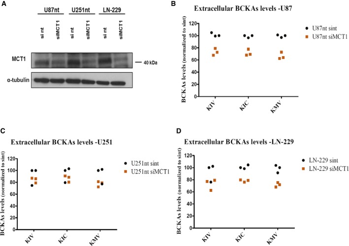
. 2017 Oct 24;18(12):2172–2185. doi: 10.15252/embr.201744154

Figure EV3. MCT1 knockdown decreases BCKA excretion.

Figure EV3

  • A
    Western blot analysis of MCT1 levels in U87‐MG cells, U251‐MG expressing normal levels of BCAT1 (U87nt and U251nt, respectively) and LN‐229 cells 48 h post‐transfection using DharmaFECT 1 transfection reagent (Dharmacon) with 25 nmol of MCT1 siRNA smart pool (Dharmacon), or non‐target (nt) #2 siRNA pool (Dharmacon). Anti‐MCT1 antibody (AB3538P, Millipore) was used at 1:5,000 dilution. α‐tubulin was used as loading control.
  • B–D
    BCKAs (KIV, KIC, KMV) levels are determined by ultra performance liquid chromatography (UPLC) coupled to fluorescence detection in supernatants from U87nt (B), U251nt (C), and LN‐229 (D) cells 48 h after transfection. BCKAs levels are normalized to total protein content and to the levels detected in U87nt, U251nt, or LN‐229 cells transfected with non‐target #2 siRNA (U87nt sint, U251nt sint, or LN‐229 sint, respectively). n = 3 technical replicates. KIV: α‐ketoisovalerate. KIC: α‐ketoisocaproate. KMV: α‐keto‐β‐methylvalerate.