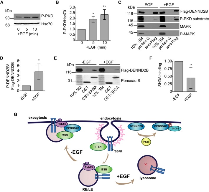
Figure 5. EGF treatment dissociates DENND2B from ITSN.

- HEK‐293T cells were serum‐starved, treated with 100 ng/ml EGF for the indicated times, and cell lysates were analysed by Western blot.
- Quantification of (A) where P‐PKD is normalized to Hsc70. Mean ± SD. ANOVA with Dunnett's post‐test *P = 0.015 and **P = 0.003, n = 3 from three independent experiments.
- HEK‐293T cells were serum‐starved, treated with 100 ng/ml EGF for 5 min, and cell lysates were incubated with protein G beads or protein G beads coupled to anti‐Flag. Bound proteins were detected by Western blot.
- Quantification of (B) where P‐PKD substrate is normalized to Flag‐DENND2B. Mean ± SD, Welch's t‐test *P = 0.036, n = 4 from three independent experiments.
- HEK‐293T cells were serum‐starved, treated with 100 ng/ml EGF for 5 min, and cell lysates were incubated with GST‐SH3A. Total proteins and bound proteins were detected by Ponceau S staining and Western blot, respectively.
- Quantification of (E) where bound Flag‐DENND2B is normalized to SM. Mean ± SD, Welch's t‐test *P = 0.012, n = 6 from three independent experiments.
- Schematic of EGFR trafficking. ITSN promotes EGFR internalization upon activation. EGF‐independent activation of EGFR: EGFR traffics on Rab13‐positive recycling or late endosomes (RE/LE) to the plasma membrane where ITSN binds the DENND2B to facilitate EGFR exocytosis. EGF‐dependent activation of EGFR: DENND2B is phosphorylated by PKD, binds 14‐3‐3 and loses its affinity for ITSN and EGFR is targeted towards lysosomal degradation.
Source data are available online for this figure.
