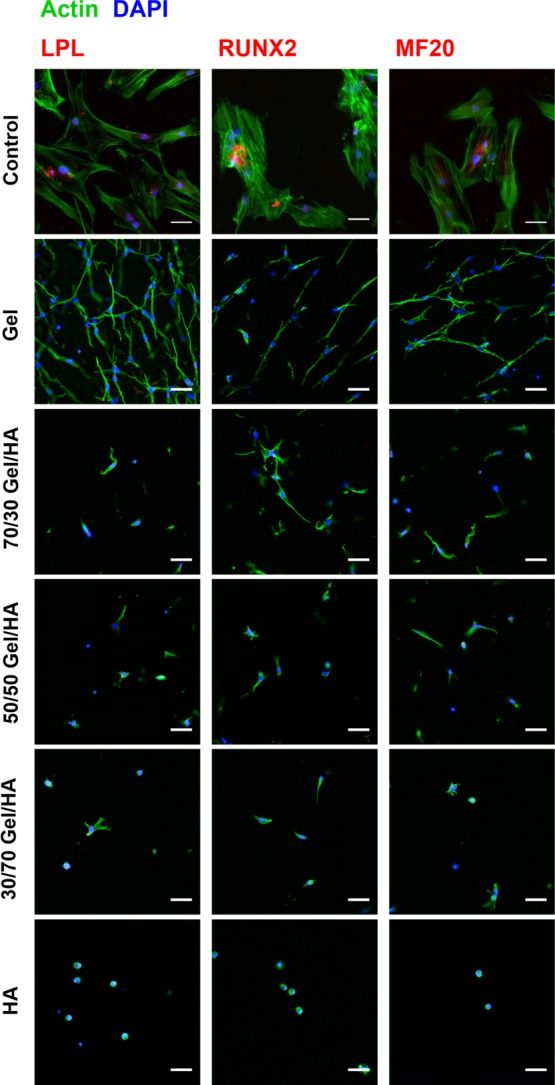
Figure 5.

Immunofluorescence images for LPL, RUNX2, and MF20 of hMSCs cultured in Gel–HA hydrogels and in the GM for 14 days. Nuclei are stained with 4′,6-diamidino-2-phenylindole (DAPI), cytoskeleton is stained in green, and the different antibodies are stained in red. The scale bar is 50 μm.
