Fig. 3.

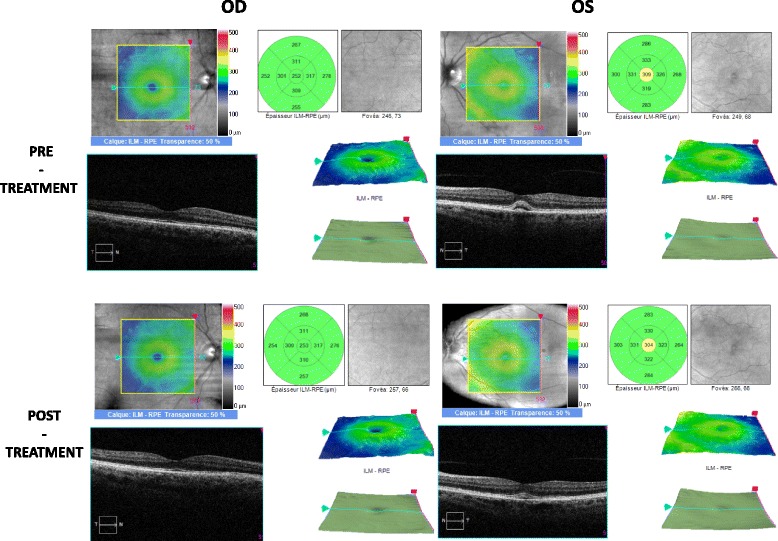
Bilateral optical coherence tomography: OD: normal foveolar profile before and after treatment, OS: retrofoveal subretinal detachment before and after treatment

Bilateral optical coherence tomography: OD: normal foveolar profile before and after treatment, OS: retrofoveal subretinal detachment before and after treatment