Fig. 1.

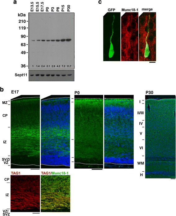
Expression of Munc18–1 in developing mouse brain. a Developmental changes of Munc18–1 protein amounts. Whole lysates (20 μg protein) of cerebral cortices at various developmental stages were subjected to western blotting (10% gel) with anti-Munc18–1. Anti-Sept11 was used for a loading control. The expression level of Munc18–1 was corrected based on that of Sept11 using ImageJ software, and relative expression was shown as fold-increase over the expression level at E13.5. b Munc18–1 distribution in developing cerebral cortex. Coronal sections were examined for Munc18–1 (green) and nuclei (blue) by immunohistochemical staining at E17, P0 and P30. Bars, 100 μm. A cortical slice (E17) was double-stained for Munc18–1 (green) and Tag-1 (red). Tag-1 distribution and a merged image were shown. Bar, 50 μm. c Subcellular distribution of Munc18–1 in migrating neurons in the CP. pCAG-GFP was electroporated into cerebral cortices at E14.5 and fixed at E17 to visualize migrating neurons. Coronal sections were prepared and stained for GFP and Munc18–1. A representative neuron in the lower CP was displayed. Bar, 5 μm
