Fig. 6.

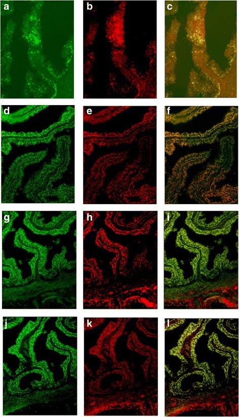
CK7-positive staining in the cytoplasm. Observed red fluorescence of CK7 overlapped with green fluorescence of WJMSCs with GFP in tubal epithelium. Yellow fluorescence of colocalization of CK7 and WJMSCs observed in all four groups. a–c Control A group. d–f Control B group. g–i Experimental C group. j–l Experimental D group
