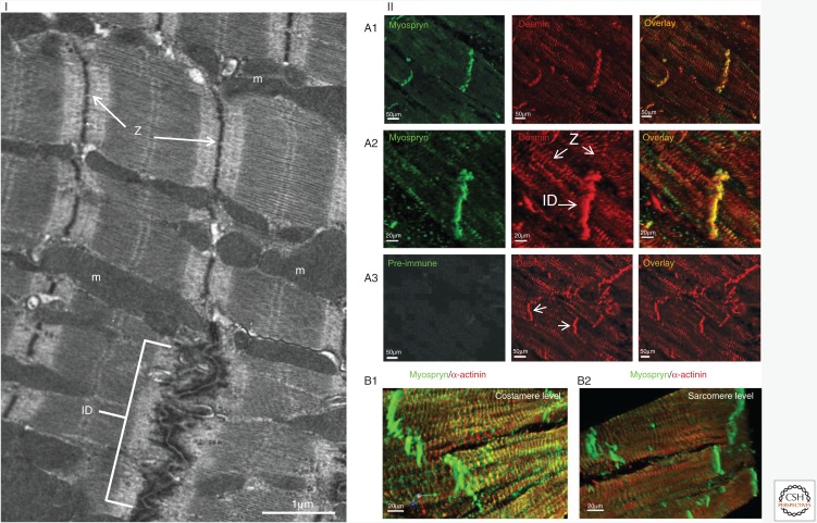
Figure 2.
Localization of desmin and its associated protein myospryn in cardiac striated muscle. (I) Electron-microscopic ultrastructural view of the mouse myocardium showing intercalated disks (ID), Z-disks (Z), and mitochondria (m). (II) Colocalization of desmin (red) and myospryn (green) by immunofluorescence microscopy. IDs and Z-disks (Z) are indicated in panel I and the middle figure of panel IIA2; IDs are also indicated in panel IIA3 (middle) by arrowheads. (B1, B2) In contrast to ID and costameres, myospryn (green) is not detectable at the internal sarcomeric Z-discs. α-actinin (red) is used as positive Z-disc marker. Not shown here, desmin is also localized at the costameres. Scale bars, 1 μm (I), 20 μm (IIA2, IIB1, IIB2), and 50 μm (IIA1, IIA3). (II, Adapted from Kouloumenta et al. 2007.)

