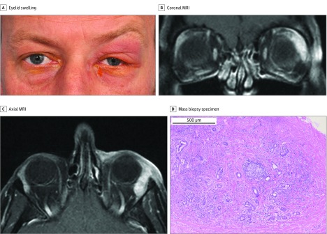
Figure 3. Idiopathic Dacryoadenitis.
A, A man in his early 40s presented with a 1-month history of tender left upper eyelid swelling. B, Coronal magnetic resonance imaging (MRI). C, Axial MRI. The MRIs demonstrated an idiopathic orbital inflammation (IOI) pattern of a well-defined, enlarged lacrimal gland with homogeneous contrast enhancement. D, Surgical biopsy specimen of the lacrimal gland mass showed IOI features with lymphoplasmacytic infiltration focally organized into a lymphoid follicle with a germinal center. Fibrosis extends into the lacrimal gland with some preserved lacrimal gland (hematoxylin-eosin, original magnification x50).

