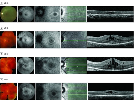
Figure 1. Composite of Retinal Imaging From the Left Eyes of Patients With Late-Onset CLN3-Associated Rod-Cone Dystrophy.
Imaging from each patient includes color fundus photograph (first panel), fundus autofluorescence (second and third panels), and en face infrared image (fourth panel) demarcating the horizontal optical coherence tomography scan line (fifth panel).

