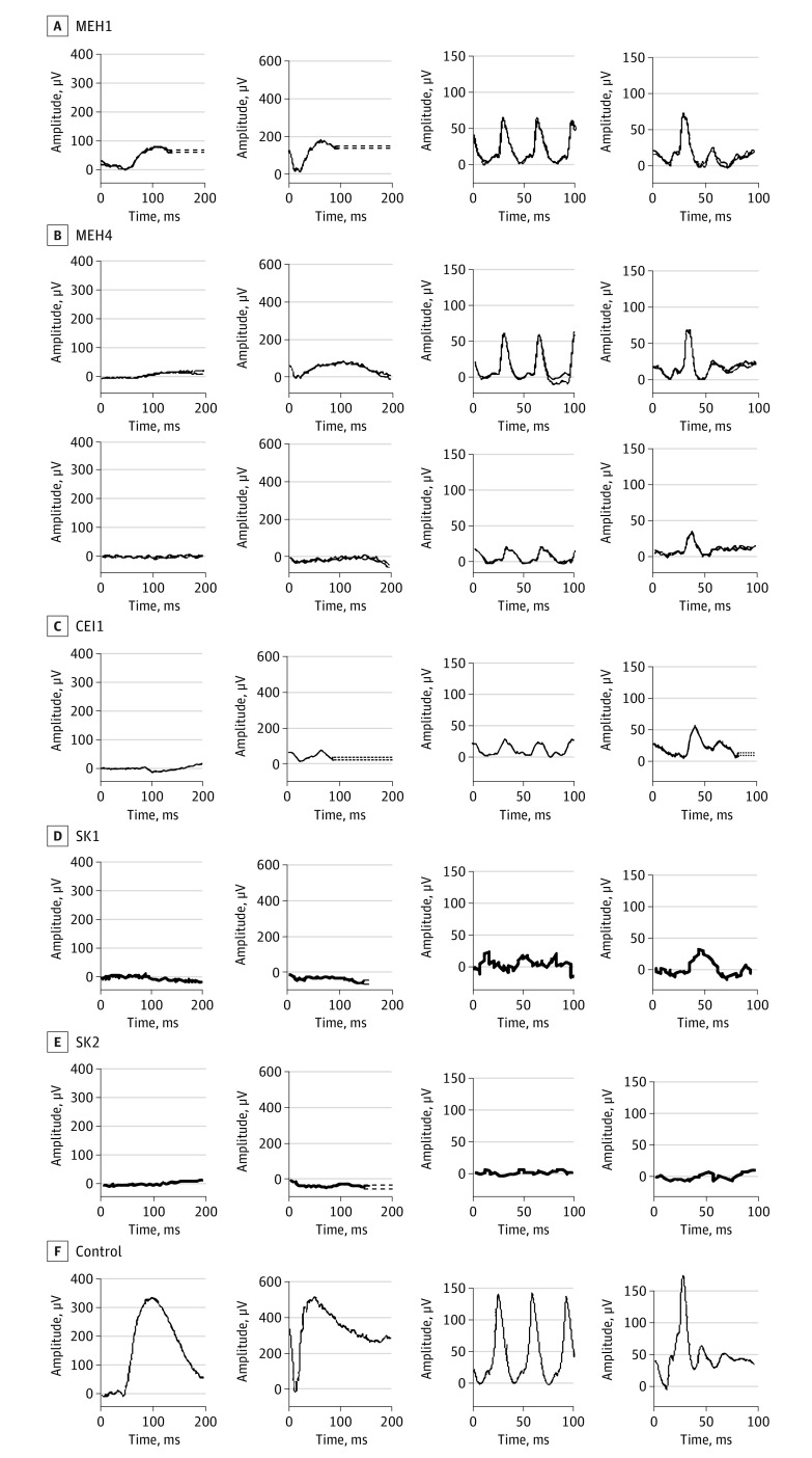
Figure 3. Full-Field Electroretinograms.
Results of full-field electroretinograms under dim scotopic (dark-adapted [DA] 0.01; first panel), bright scotopic (DA 10.0; second panel), photopic flicker (light-adapted [LA] 3.0, 30 Hz; third panel), and photopic single flash (LA 3.0; fourth panel) intensities. Results for patient MEH4 shown from testing performed in his late 30s and 5 years later.

