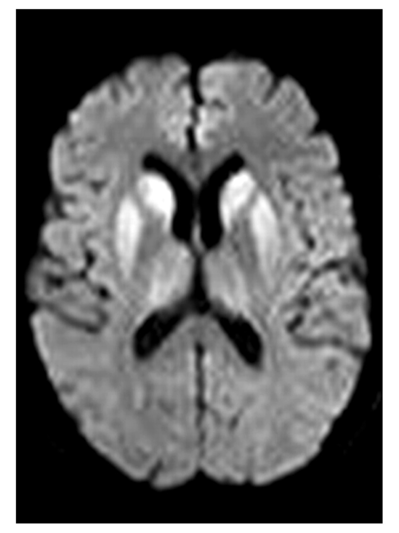
Figure 1. Magnetic resonance imaging of sporadic Creutzfeldt-Jakob disease.
Diffusion-weighted image at the level of the basal ganglia demonstrates marked symmetrical hyperintensity in the caudate head and putamen with less marked affection of the thalami. Image courtesy of David Summers, Western General Hospital, Edinburgh, UK.

