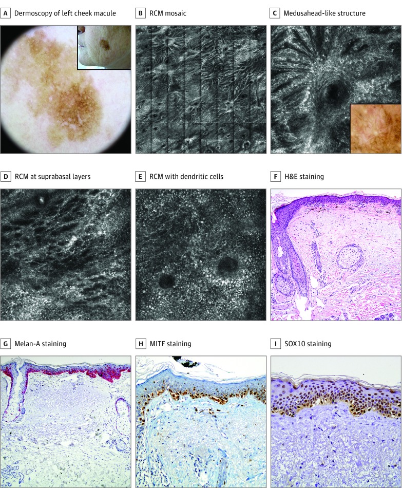
Figure 3. Lentigo Maligna in a Woman in Her 60s: False-Negative According to the Reflectance Confocal Microscopy (RCM) Algorithm.
A, An 11-mm, brown macule on the left cheek treated 6 times with cryotherapy (inset). Dermoscopically, it showed a brown pseudonetwork, pigmented rhomboidal structures, and targetlike patterns. B, An RCM mosaic (4 x 4 mm) showed an abundant number of follicular openings with radiating refractile strands composed by confluent, elongated clusters of atypical cells all over the circumference. C, An RCM image (0.5 × 0.5 mm). Detail of medusahead-like structure. Inset: correlation with dermoscopy. D, An RCM image (0.5 × 0.5 mm). Epidermal disarray with abundant dendritic pagetoid cells at suprabasal layers. E, An RCM image (0.5 × 0.5 mm). Dendritic cells with perifollicular distribution. F, Biopsy specimen revealed an atypical proliferation of melanocytes with extension along the follicular structures. Slight melanophagia and severe elastosis in the dermis. Hematoxylin-eosin (H&E), original magnification ×100. G, Melan-A staining, original magnification ×100, labeling the atypical hyperplasia of melanocytes in epidermis and adnexa. Isolated pagetoid melanocytes are visualized. H, Microphthalmia-associated transcription factor (MITF) staining, original magnification ×100, labeling the nucleus of atypical melanocytes. I, SRY-related HMG (high mobility group)-box gene 10 (SOX10) staining, original magnification ×100, expressing the same features as MITF.

