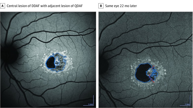
Figure 1. Examples of Progression of Lesion of Definitely Decreased Autofluorescence (DDAF) and Questionably Decreased Autofluorescence (QDAF).
A, Central lesion of DDAF (0.82 mm2) with a temporal adjacent lesion of QDAF (1.53 mm2). B, Same eye after 22 months: the lesion of DDAF is enlarged (2.09 mm2), whereas the lesion of QDAF (0.45 mm2) is decreased, indicating the transformation from QDAF into DDAF; the total area enlarged from 2.35 to 2.54 mm2.

