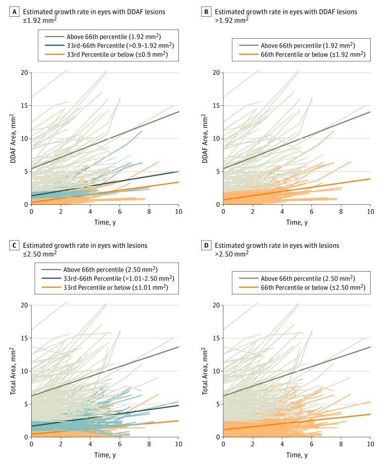
Figure 2. Estimated Yearly Progression of Areas Based on Lesion Size at the First Included Visit.
A, Estimated growth rate in eyes with definitely decreased autofluorescence (DDAF) lesion sizes ≤1.92 mm2 (66th percentile) was 0.32 mm2/y (95% CI, 0.24-0.39 mm2/y) (brown line). B, Estimated growth rate in eyes with DDAF lesion sizes >1.92 mm2 was estimated as 0.86 mm2/y (95% CI, 0.67-1.06 mm2/y) (orange line). C, For total area of decreased areas of autofluorescence, eyes with lesion sizes ≤2.50 mm2 (66th percentile) showed an estimated growth rate of 0.26 mm2/y (95% CI, 0.21-0.32 mm2/y) (brown line). D, For total area of decreased areas of autofluorescence, eyes with lesions sizes >2.50 mm2 showed an estimated growth rate of 0.74 mm2/y (95% CI, 0.57-0.91 mm2/y) (orange line). Diagonal lines in parts A-D indicate estimated growth rates.

