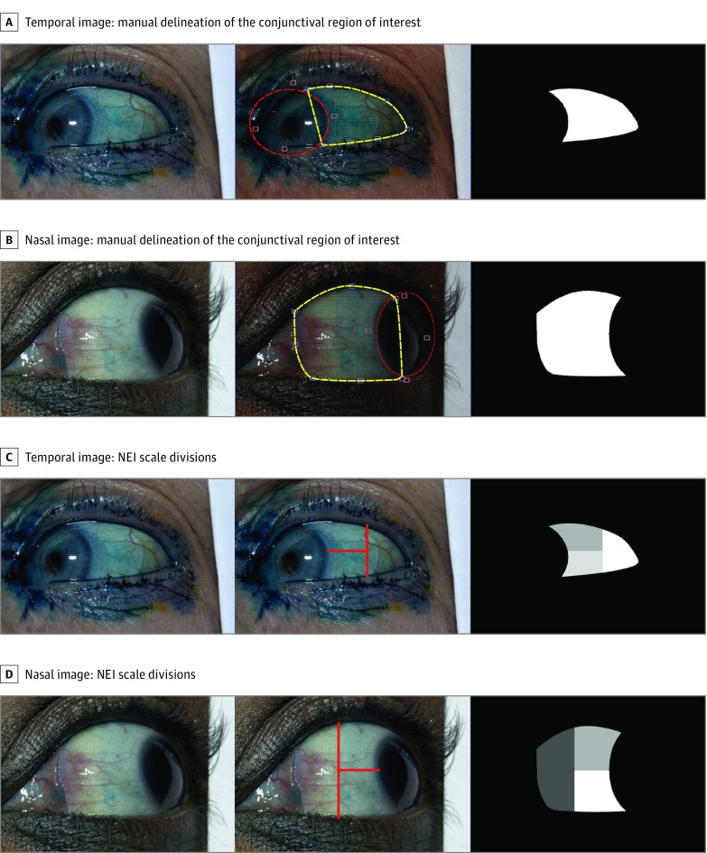
Figure 1. Examples of Manual Delineation of the Conjunctival Region of Interest and Lines Corresponding to the Divisions of the National Eye Institute (NEI) Scale for Temporal and Nasal Images.
The yellow landmarks and curves show the initial delineation of the conjunctival boundaries shown in the central panels of A and B. The red ellipse (controlled by 4 adjacent red points) is used to remove the regions of the cornea from the yellow delineation. Examples of lissamine green images with NEI lines drawn on the temporal (C) and nasal (D) image and conjunctival region of interest. The 2 delineations are combined to create the final conjunctival region of interest shown in the rightmost panels.

