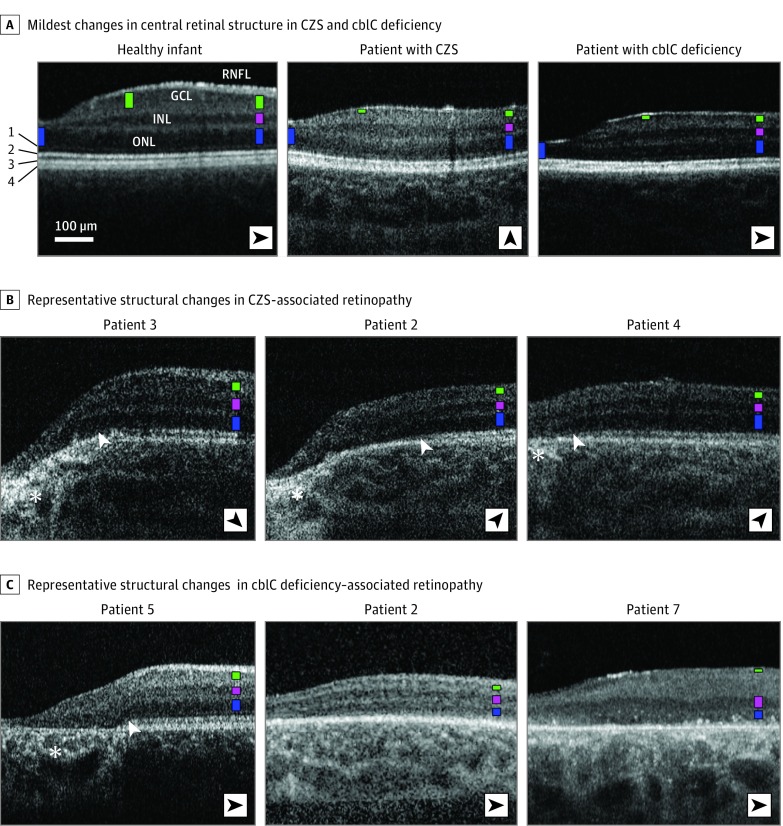
Figure 1. Spectral-Domain Optical Coherence Tomography (SD-OCT) Findings in Study Participants.
A, Nonstraightened, magnified, SD-OCT cross-sections extending 2.7 mm from the foveal center in patients with the mildest retinal abnormalities in congenital Zika syndrome (CZS)– and cobalamin C (cblC) deficiency–associated retinopathies compared with a healthy infant. Patients’ identification corresponds to that used in recent articles; fundus images have been published in Ventura et al and Bonafede et al. Nuclear layers are labeled in the healthy infant; structures distal to the outer nuclear layer (ONL) are numbered to the left as external limiting membrane (1), inner-outer segment boundary zone or ellipsoid zone (2), interdigitation between the tip of the photoreceptors and the apical retinal pigment epithelium (RPE; 3), and RPE (4). To visualize comparisons in thicknesses of the different nuclear layers, colored bars are overlaid on each nuclear layer at 2 mm of eccentricity (ONL, blue; inner nuclear layer [INL], pink; and ganglion cell layer [GCL], green) and at locations where maximal thickness values for ONL (foveal center) and GCL thickness (approximately 1 mm from the foveal center) are approached. Orientation of the scans varied (black arrowheads). B and C, Examples of cross-sections in patients with CZS– and cblC deficiency–associated retinopathy. White arrowheads indicate regions of transition with loss of the ellipsoid zone signal. Asterisks mark increased posterior backscattering from RPE demelanization or loss. RNFL indicates retinal nerve fiber layer.

