Key Points
Question
For patients suspected of having emergent large-vessel occlusion (ELVO) who present to a primary stroke center (PSC), can a standardized protocol that is based on (1) early notification to the closest comprehensive stroke center (CSC), (2) computed tomographic angiography at the PSC, and (3) electronic image sharing prior to transfer improve efficiency and outcomes?
Findings
In this cohort study, when the PSC protocol was fully executed, the rate of good outcomes was doubled and the time from arrival at the PSC to reperfusion at the CSC was almost 1 hour less than that with only a partial execution of the protocol.
Meaning
This protocol can be easily replicated between PSC and CSC partners and may improve stroke care delivery for patients with ELVO presenting to centers without endovascular capability.
Abstract
Importance
While prehospital triage to the closest comprehensive stroke center (CSC) may improve the delivery of care for patients with suspected emergent large-vessel occlusion (ELVO), efficient systems of care must also exist for patients with ELVO who first present to a primary stroke center (PSC).
Objective
To describe the association of a PSC protocol focused on 3 key steps (early CSC notification based on clinical severity, vessel imaging at the PSC, and cloud-based image sharing) with the efficiency of care and the outcomes of patients with suspected ELVO who first present to a PSC.
Design, Setting, and Participants
In this retrospective cohort study, 14 regional PSCs unfamiliar with the management of patients with ELVO were instructed on the use of the following protocol for patients presenting with a Los Angeles Motor Scale score 4 or higher: (1) notify the CSC on arrival, (2) perform computed tomographic angiography concurrently with noncontract computed tomography of the brain and within 30 minutes of arrival, and (3) share imaging data with the CSC using a cloud-based platform. A total of 101 patients were transferred from regional PSCs to the CSC between July 1, 2015, and May 31, 2016, and received mechanical thrombectomy for acute ischemic stroke. The CSC serves approximately 1.7 million people and partners with 14 PSCs located between 6.4 and 73.6 km away. All consecutive patients with internal carotid artery or middle cerebral artery occlusions transferred over an 11-month period were reviewed, and they were divided into 2 groups based on whether the PSC protocol was partially or fully executed.
Main Outcomes and Measures
The primary outcomes were efficiency measures including time from PSC door in to PSC door out, time from PSC door to CSC groin puncture, and 90-day modified Rankin Scale score (range, 0-6; scores of 0-2 indicate a good outcome).
Results
Although 101 patients were transferred, only 70 patients met the inclusion criteria during the study period. The protocol was partially executed for 48 patients (68.6%) (mean age, 77 years [interquartile range, 65-84 years]; 22 of the 48 patients [45.0%] were women) and fully executed for 22 patients (31.4%) (mean age, 76 years [interquartile range, 59-86 years]; 13 of the 22 patients [59.1%] were women). When fully executed, the protocol was associated with a reduction in the median time for PSC arrival to CSC groin puncture (from 151 minutes [95% CI, 141-166 minutes] to 111 minutes [95% CI, 88-130 minutes]; P < .001). This was primarily related to an improvement in the time from PSC door in to door out that reduced from a median time of 104 minutes (95% CI, 82-112 minutes) to a median time of 64 minutes (95% CI, 51-71.0 minutes) (P < .001). When the protocol was fully executed, patients were twice as likely to have a favorable outcome (50% vs 25%, P < .04).
Conclusions and Relevance
When fully implemented, a standardized protocol at PSCs for patients with suspected ELVO consisting of early CSC notification, computed tomographic angiography on arrival to the PSC, and cloud-based image sharing is associated with a reduction in time to groin puncture and improved outcomes.
This cohort study reports on the association of a primary stroke center protocol with the efficiency of care and the outcomes of patients with suspected emergent large-vessel occlusion who first present to a primary stroke center.
Introduction
In addition to intravenous (IV) tissue plasminogen activator (tPA) for eligible patients, endovascular treatment for acute ischemic stroke caused by emergent large-vessel occlusion (ELVO) in the anterior circulation is now the standard of care for patients presenting within 6 hours of symptom onset. Since the treatment effect of mechanical thrombectomy for stroke by LVO is time dependent, the innovation of the delivery of care to patients with stroke is now a major focus, with systems reorganization and timely access being top priorities.
While prehospital triage to the closest comprehensive stroke center (CSC) may be the most efficient means to improve the delivery of stroke care for patients with ELVO, effective systems of care must also remain in place for patients with ELVO presenting to a primary stroke center (PSC). For this reason, we created a process focused on early CSC notification, obtaining vessel imaging at the PSC, and image sharing, without a formal telestroke relationship. The aim of this article is to outline this process and report the outcomes observed after implementation of this PSC ELVO protocol.
Methods
Study Design, Setting, and Participants
Lifespan’s institutional review board, a committee made up of physicians, scientists, and community members who ensure that research involving human subjects is well planned and ethical, approved this study, which retrospectively reviewed data from our prospectively collected stroke center quality assurance database. Informed consent was waived because the data were deidentified. Our CSC serves a population of nearly 1.7 million people and is the closest CSC to 14 PSCs and 3 stroke-ready hospitals. We included all patients transferred from regional PSCs to our CSC between July 1, 2015, and May 31, 2016, who received mechanical thrombectomy for acute ischemic stroke. The starting date was chosen based on when the new process was first introduced, and time metrics associated with the PSC to CSC transfers were tracked (no historical data for these patients exists because there was no precedent for identifying and transferring these patients). Thrombectomy candidacy criteria were based on the most recent American Heart Association guidelines. All patients were transported directly to our angiography suite, and those who underwent attempted mechanical thrombectomy were included in the study. Conscious sedation was used in all patients except those who had been intubated at the PSC (n = 2).
Study Treatment and Intervention
Patients were divided into 2 groups based on whether the PSC ELVO protocol was partially or fully executed.
PSC ELVO Protocol
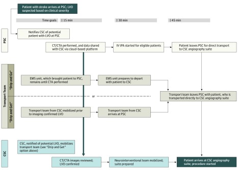
We standardized a process for the rapid diagnosis and transfer of patients with ELVO presenting to PSCs. The key steps to this process are as follows:
Emergency department physicians assess patients with potential stroke immediately on arrival to the PSC and call our CSC if the patient’s Los Angeles Motor Scale (LAMS) score is 4 or higher. This is before any imaging has been performed.
Following this telephone call from the emergency department physicians, the CSC critical care transport team is dispatched. If the CSC transport team is unavailable, the PSC can make alternate arrangements to have a transport team ready prior to confirmation of LVO by computed tomographic angiography (CTA). The mantra here is “waste gas not brain.”
If the LAMS score is 4 or higher, the PSC performs CTA at the time of initial noncontrast computed tomography (NCCT) of the brain, and ideally within 30 minutes of PSC arrival.
The images are sent via a secure, Health Insurance Portability and Accountability Act–compliant cloud-based platform for remote viewing by the CSC stroke team.
All patients with confirmed ELVO are directly transported to the CSC angiography suite.
The critical aspects of each of these steps are described in greater detail in Figure 1 and in the eAppendix in the Supplement. Patients for whom all PSC ELVO protocol steps were followed were classified as being in the “full execution” group. Patients for whom some, but not all, steps were followed were classified as being in the “partial execution” group (see eTable 1 in the Supplement for greater detail of partial execution group).
Figure 1. Primary Stroke Center (PSC) Emergent Large-Vessel Occlusion (ELVO) Protocol.
The PSC ELVO protocol workflow is focused on early notification of the comprehensive stroke center (CSC; with a goal of <30 minutes), obtaining early vessel imaging (with a goal of <30 minutes) at the PSC, and cloud-based image sharing. CTA indicates computed tomographic angiography of the brain and neck; CT, computed tomography of the brain; EMS, emergency medical services; and IV tPA, intravenous tissue plasminogen activator (alteplase).
Demographics, Variable, and Measurements
The demographics of interest include age, sex, originating PSC, admission National Institute of Health Stroke Scale (NIHSS) score, premorbid modified Rankin Scale (mRS) score, site of intracranial occlusion, NCCT Alberta Stroke Program Early Computed Tomography Score, distance between PSC and CSC. The 4 PSCs located more than 40 km (25 miles) driving distance from the CSC were considered “long-distance” transport, and the remaining were considered “short-distance” transport. All patients were transported using ground transport.
The time intervals of interest included the time from onset to PSC door, the time from PSC arrival to treatment with IV tPA, the time from PSC NCCT to CSC groin puncture, the time from PSC arrival to PSC departure (door in to door out [DIDO]), the time from PSC arrival to CSC groin puncture, the time from PSC arrival to CSC recanalization, the time from CSC arrival to CSC groin puncture, and recanalization time. Recanalization was defined as the time of first angiographic image showing a modified thrombolysis in cerebral infarction score of 2b, 2c, or 3 in the territory of interest. Compliance with the protocol (partial vs complete) and workflow metrics on a per-center basis are also reported in eTable 2 in the Supplement.
Outcome metrics included discharge NIHSS and mRS score at 90 days. A 90-day mRS score of 0 to 2 was defined as a favorable outcome.
Statistical Analysis
All analyses were conducted using SAS software version 9.4 (SAS Inc). Demographics were examined for counts with differences being examined with the χ2 or Fisher exact test; ordinal and continuous variables were examined (median, first quarter to third quarter, and minimum and maximum values [range]), and differences were assessed using a Wilcoxon test. The time between events was estimated using the Kaplan-Meier estimation (median values and 95% CIs), and differences between PSC protocol and no protocol were assessed using a Wilcoxon test, given that no observations were censored. Modeling outcomes include the mRS score, between initial arrival and at 90 days, along with NIHSS, between admission and discharge, which were examined by full and partial PSC protocol, using generalized mixed modeling with the classic sandwich estimation; this modeling was performed by assuming a binominal distribution (0-6 and 0-42, respectively). Potential moderators were the distance between the PSC and the CSC, time from ELVO onset to treatment with tPA, time from door to needle, CSC notified in 30 minutes, CTA at PSC in 30 minutes, image availability on lifeIMAGE at http://www.lifeimage.com/, and successful reperfusion; however, no significant effects were observed for these moderators. Statistical significance was established a priori at the .05 level, and all interval estimates were calculated for 95% confidence. Multiple comparisons were examined using the Tukey method. For clinical outcomes, reported mean values are not arithmetic or continuous but are derived from a logit link and binomial space, then back-transformed into the original scale value.
Results
A total of 101 patients with confirmed (CTA positive at PSC) ELVO and last seen well within 6 hours were transported to our CSC for thrombectomy during the study period. Of these, 70 patients with angiographically confirmed internal carotid artery and/or middle cerebral artery (M1 segment) occlusions who underwent attempted mechanical thrombectomy were included in the analysis. Among the 31 patients excluded, there were 10 patients with clot lysis during transport (confirmed by angiography), 10 patients with isolated M2 occlusions, 7 patients with posterior circulation occlusions, 3 patients who were inpatients at the outside hospital (ie, not presenting to the PSC emergency department), and 1 patient who arrived at our institution, where treatment was deferred, with an NIHSS score of 0.
The distances from which the patients with ELVO were transferred ranged from 6.4 to 75.2 km (4 to 47 miles). Thirty-one patients (44.3%) were transported from 4 hospitals more than 40.0 km (25 miles) away (long distance) and 39 patients (55.7%) were transported from hospitals 40.0 km (25 miles) away or less (short distance). Sixty-six patients (94.3%) underwent CTA at the PSC. The other 4 patients underwent CTA on arrival to the CSC because it had not been performed at the time of the initial NCCT and would have delayed transportation to our CSC. The PSC ELVO protocol was fully executed for 22 of the 70 patients (31.4%) and either partially or not executed for 48 of the 70 patients (68.6%) (Table 1), and there were no differences between the 2 groups with regard to age, NIHSS score, ASPECTS, sex, IV tPA administration, and successful reperfusion (modified thrombolysis in cerebral infarction score of 2b, 2c, or 3). There was 1 symptomatic hemorrhage in the study group, and this occurred in a patient who received IV tPA and for whom the PSC protocol was fully executed (the time from PSC door to CSC recanalization was 100 minutes).
Table 1. Demographic and Biometric Outcomes When the PSC Protocol Was Partially or Fully Executed.
| Data | Patients in Partial Protocol Group (n = 48) |
Patients in Full Protocol Group (n = 22) |
P Value |
|---|---|---|---|
| Age, y | |||
| Median (Q1-Q3) | 77 (65-84) | 75.5 (59-86) | .70 |
| Range | 33-98 | 19-92 | |
| Female, No. (%) | 22 (49) | 13 (59) | .30 |
| Initial NIHSS score | |||
| Median (Q1-Q3) | 19 (13-22) | 17 (15-22) | .47 |
| Range | 6-30 | 9-32 | |
| NCCT ASPECTS | |||
| Median (Q1-Q3) | 10 (8-10) | 10 (9.5-10) | .20 |
| Range | 5-10 | 7-10 | |
| Long distance, No. (%)a | 20 (41.7) | 11 (50.0) | .51 |
| Distance, median (range), kmb | 32.2 (6.4-73.6) | 40.8 (7.0-73.6) | .45 |
| IV tPA administered, No. (%) | 31 (64.6) | 14 (63.6) | .94 |
| Outcomes, No. (%) | |||
| Successful reperfusionc | 38 (79.2) | 21 (95.5) | .15 |
| Favorable outcome at 90 dd | 12 (25.0) | 11 (50.0) | .04 |
Abbreviations: ASPECTS, Alberta Stroke Program Early Computed Tomography Score; IV tPA, intravenous tissue plasminogen activator (alteplase); NCCT, noncontract computed tomography; NIHSS, National Institutes of Health Stroke Scale; PSC, primary stroke center; Q1-Q3, first quarter to third quarter.
More than 40 km (25 miles) from the comprehensive stroke center.
To convert kilometers to miles, divide by 1.6.
Modified thrombolysis in cerebral infarction score of 2b, 2c, or 3.
Modified Rankin Scale score of 0 to 2.
Differences in Care Efficiency Metrics
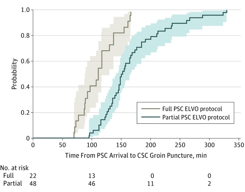
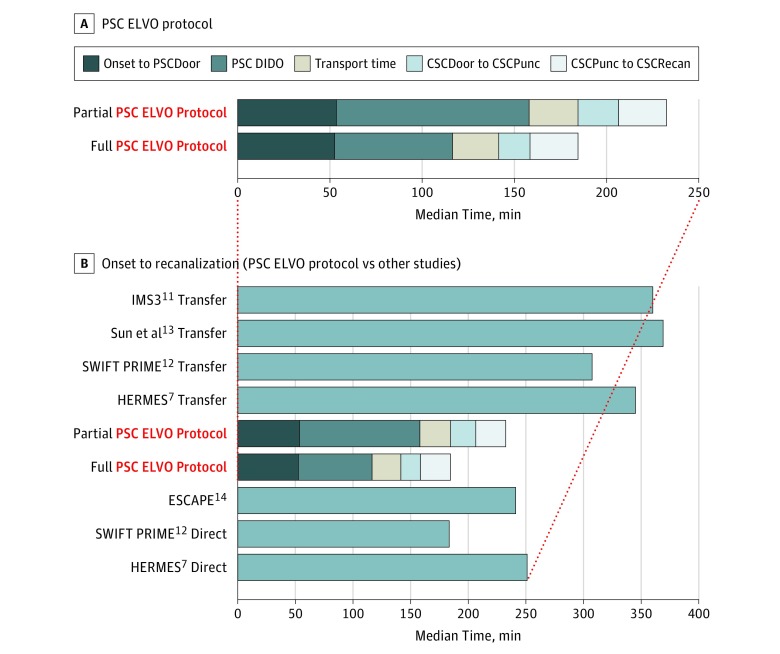
As indicated in Table 2, the workflow metrics were significantly faster when the PSC protocol was fully executed. Specifically, the median duration between PSC arrival and CSC groin puncture was 111 minutes when fully executed, 40 minutes less than when partially executed (P < .001) (Figure 2). Relatedly, the median duration from door in to door out was 64 minutes when the protocol was executed fully and 104 minutes when partially executed (P < .001). Also of note is the reduction in door-to-needle times for IV tPA at PSCs when the protocol was fully executed relative to partially executed (39.5 vs 65 minutes; P < .001). No difference was observed in the median recanalization time between the 2 groups (ie, 26 minutes for both groups; P = .82). Figure 3A graphically depicts the key time intervals; improvement in onset to recanalization appears to be driven by improved inhospital processes at the PSC translating to decreased DIDO times. Figure 3B graphically depicts care efficiency with other reported series of patients with ELVO who were either transferred or directly admitted to a CSC (endovascular-capable center).
Table 2. Differences in Workflow Metrics Between Partially and Fully Executed PSC Protocol.
| Metric | Partial Protocol | Full Protocol | P Value | ||
|---|---|---|---|---|---|
| Median (IQR) | 95% CI | Median (IQR) | 95% CI | ||
| PSC | |||||
| OTD | 53.5 (37-118) | 45.0-67.0 | 52.5 (32-129) | 32.0-79.0 | .64 |
| DTN | 65.0 (55-96) | 58.0-78.0 | 39.5 (32-51) | 30.0-51.0 | <.001 |
| Onset to IV tPA | 113.0 (92-165) | 100.0-125.0 | 92.0 (60-112) | 60.0-112.0 | .01 |
| CSC | |||||
| OTPunc | 218.5 (176-326) | 191.0-255.0 | 185.0 (137-209) | 137.0-205.0 | .02 |
| OTRep | 262.0 (203-372) | 223.0-316.0 | 222.0 (165-275) | 165.0-263.0 | .04 |
| PSC NCCT to CSCPunc | 133.0 (108-165) | 120.0-139.0 | 97.5 (76-120) | 76.0-110.0 | <.001 |
| PSC CTA to CSCPunc | 110.5 (79-136) | 95.0-122.0 | 93.0 (69-122) | 69.0-111.0 | .16 |
| DIDO PSC | 104.5 (78-121) | 82.0-112.0 | 64.0 (51-88) | 51.0-71.0 | <.001 |
| PSCDoor to CSCPunc | 151.0 (132-187) | 141.0-166.0 | 111.0 (88-137) | 88.0-130.0 | <.001 |
| PSCDoor to CSCRep | 179.0 (159-237) | 164.0-210.0 | 132.0 (113-183) | 113.0-172.0 | <.001 |
| CSCDoor to CSCPunc | 22.0 (17-32) | 19.0-26.0 | 17.0 (14-20) | 14.0-20.0 | .005 |
| CSC reperfusion time | 26.0 (15-41) | 19.0-32.0 | 26.0 (15-30) | 15.0-30.0 | .82 |
Abbreviations: CSC, comprehensive stroke center; CSCPunc, CSC groin puncture; CSCRep, CSC reperfusion; CTA, computed tomographic angiography of brain and neck; DIDO, door in to door out; DTN, door to needle; IQR, interquartile range; IV tPA, intravenous tissue plasminogen activator (alteplase); NCCT, noncontrast computed tomography of brain; OTD, onset to door; OTPunc, onset to groin puncture; OTRep, onset to reperfusion; PSC, primary stroke center; PSCDoor, arrival at PSC.
Figure 2. Primary Stroke Center (PSC) Emergent Large-Vessel Occlusion (ELVO) Protocol Efficiency From PSC Arrival to Comprehensive Stroke Center (CSC) Groin Puncture.
Kaplan-Meier time-to-event curves with 95% CIs, reflecting transfer efficiency achieved when the PSC ELVO protocol was fully executed vs when it was partially executed. As demonstrated by the steep growth of the grey curve, 9 patients in the fully executed group had a time from PSC arrival to CSC groin puncture of less than 100 minutes. Note that the steep growth of the grey curve implies both greater overall speed and less variability.
Figure 3. Primary Stroke Center (PSC) Emergent Large-Vessel Occlusion (ELVO) Protocol Care Efficiency Metrics.
A, Graphical depiction of key time intervals from onset to reperfusion for a PSC ELVO protocol that is partially vs fully executed. Note that the primary driver of improvement in onset-to-reperfusion times appears to be reduction in time from PSC arrival to PSC departure (from door in to door out [DIDO]). B, Comparison of the partially and fully executed PSC ELVO protocols with other series of patients with ELVO who were either transferred from a PSC or directly admitted to a comprehensive stroke center (CSC). CSCDoor indicates arrival at CSC; CSCPunc, CSC groin puncture; CSCRecan, CSC recanalization; ESCAPE, Endovascular Treatment for Small Core and Proximal Occlusion Ischemic Stroke; HERMES, Highly Effective Reperfusion Evaluated in Multiple Endovascular Stroke Trials; IMS3, Interventional Management of Stroke III; PSCDoor, arrival at PSC; SWIFT PRIME, Solitaire with the Intention for Thrombectomy as Primary Endovascular Treatment for Acute Ischemic Stroke.
Clinical Outcomes
NIHSS Score at Discharge
Among those patients for whom the PSC protocol was partially executed, the median (and mean [95% CI]) admission NIHSS score was 19 (17.5 [95% CI, 15.1-19.9]), and the median (and mean [95% CI]) discharge NIHSS score was 5.5 (9.5 [95% CI, 7.8-11.4]), a reduction of 46%. Among those patients for whom the PSC protocol was fully executed, the median (and mean [95% CI]) admission NIHSS score was 17 (18.7 [95% CI, 15.1-22.4]), and the median (and mean [95% CI]) discharge NIHSS score was 4.5 (6.8 [95% CI, 4.9-9.2]), a reduction of 64%. Thus, the reduction in NIHSS score with full PSC protocol execution was significantly greater than that with incomplete or no execution (64% vs 46%) (interaction effect, P < .001).
Dichotomized mRS Score (0-2 vs 3-6)
Overall, 23 patients (32.9%) had a favorable outcome at 90 days. However, the good outcome rate was 50% in the fully executed group vs 25% in the partially executed group (P = .04). Thus, the odds of a favorable outcome increased 3 times with full PSC protocol execution relative to partial execution (odds ratio, 2.99 [95% CI, 1.0-8.7]) (Table 1).
mRS Score Distribution (0-6)
No difference was observed in premorbid mRS scores between patients for whom the PSC protocol was not executed (mRS score of 1.0 [95% CI, 0.7-1.5]) and patients for whom the PSC protocol was fully executed (mRS score of 0.6 [95% CI, 0.3-1.1]) (P = .42). However, the 90-day mRS scores were significantly lower for those for whom the PSC protocol was fully executed (mRS score of 2.3 [95% CI, 1.4-3.3]) relative to those for whom it was partially executed (mRS score of 4.1 [95% CI, 3.4-4.7]) (P = .03).
Discussion
In this series, we report the workflow metrics and clinical outcomes of a PSC protocol focused on early CSC notification, CTA acquisition at the PSC initially evaluating the patient, and cloud-based image sharing for remote CSC review prior to transfer. When fully implemented, this protocol was associated with shorter PSC DIDO times, faster times from PSC arrival to CSC recanalization, and improved outcomes. In addition, the times from PSC arrival to CSC recanalization can rival and even exceed those achieved for patients who directly present to a CSC (Figure 3B) in some series. It is worth noting that the entire spectrum of stroke care delivery metrics improved (Table 2), including a reduction from 65 to 39.5 minutes in the time from PSC arrival to IV tPA (time from PSC door to needle). Of note, only 2 of the 14 PSCs are part of our hospital system, and patients from these PSCs represent just 11% of the patient sample.
Time has a profound effect on outcomes for patients with ELVO. The investigators of the Multicenter Randomized Clinical Trial of Endovascular Treatment of Acute Ischemic Stroke in the Netherlands found a strong inverse relationship between time to reperfusion and outcome in their cohort of patients with ELVO, and they determined that the absolute difference for a good outcome is reduced by 6% per hour of delay. Similarly, the investigators of the Highly Effective Reperfusion Evaluated in Multiple Endovascular Stroke Trials recently reported that for every 15-minute acceleration time in onset to treatment, at least 39 more patients (out of 1000) will be functionally independent at 90 days. Therefore, it is of paramount importance to maximize the efficiency of the transfer process for patients with ELVO, which revolves around hospital processes at PSCs. In our study, full implementation of the protocol reduced the time from PSC arrival to recanalization by nearly 50 minutes (Table 2).
Among previous series examining similar transfer networks, Sun et al showed markedly prolonged times from initial CT to groin puncture for transfer patients compared with patients directly admitted to the “mothership” (directly to endovascular-capable centers). Their initial median time from NCCT to CSC groin puncture was 205 minutes (interquartile range [IQR], 163-274 minutes), and our initial median time was 97.5 minutes (IQR, 76-110 minutes) in the fully executed PSC protocol group. In their series, the time from PSC CT to CSC notification was a median of 66 minutes, which is longer than the median of DIDO times for our patients when the protocol was fully executed. This suggests that much of the inefficiency in transfers revolves around hospital processes at PSCs. Their data also suggested that obtaining vessel imaging at the PSC delayed the transfer process, but our results would argue against that. It is likely that, in their case, the CTA was performed in a delayed fashion, as opposed to on arrival and in conjunction with the NCCT, as our protocol calls for.
Another recent publication by Liang et al examining emergency department transfers showed no opportunity cost in outside hospital arrival to groin puncture for those patients who received vessel imaging at the outside hospital (PSC) vs the CSC. However, our median times from door in to door out were lower, at 64 minutes (fully executed) or 104.5 minutes (partially executed), compared with 142 minutes in their series. Another recent study examined a cohort of 941 patients, 124 directly admitted and 817 referred from PSC for thrombectomy over a 5-year span. Their protocol, similar to ours, did not include any repeated imaging prior to intervention at the CSC. They found a median time from PSC imaging to groin puncture of 150 minutes for transferred patients compared with a median time of 97.5 minutes for our fully executed group. Favorable outcome rates were 50% in our fully executed group and 36.4% for their series.
Obtaining vessel imaging at the PSC may improve efficiency on arrival to the CSC compared with recent SWIFT PRIME transfer data. In our series, the median time from CSC arrival to groin puncture was 17 minutes (IQR, 14-20 minutes), and the median time from CSC arrival to recanalization was 42 minutes (IQR, 29-50 minutes). These times are 46 and 62 minutes shorter, respectively, than those achieved in the SWIFT PRIME study, in which the median time from CSC arrival to groin puncture was 63 minutes (IQR, 52-81 minutes), and the median time from CSC arrival to recanalization was 104 minutes (IQR, 86-137 minutes). Certainly, it is possible that the clinical trial enrollment and randomization process may have extended the workflow metrics in the previously described trials.
There is still room for improvement in this process. Early notification of the CSC within 30 minutes of PSC arrival happened for only 44% of patients, with only 10 notifications (14%) occurring within 15 minutes (our new target). The DIDO metric is a good measure of PSC ELVO protocol efficiency as a whole, and we have set 45 minutes as our new goal for our PSC partners. Future educational initiatives for emergency department physicians, as well as other staff, should help reduce variability. In addition, one can imagine a future scenario where an emergency medical services unit transporting a patient with high clinical suspicion for ELVO to a PSC can notify the PSC prior to arrival, and that unit remains until either vessel imaging is performed or an alternate transport team can be mobilized. As soon as vessel imaging confirms ELVO and intravenous tPA is initiated, the patient can continue to the CSC. This paradigm is more akin to “drip and go” (with or without tPA) rather than “drip and ship.”
Limitations
This study has several limitations. First, this scenario might not be as applicable in other geographic areas, where the topography and population density might preclude rapid transfers. However, several aspects of this process can still apply. Mobilization of a ground transport team (air transport may be cost-prohibitive) can occur based on clinical suspicion, and vessel imaging can still be performed at the PSC, while the transport team is en route. Second, while we advocated LAMS as the screening examination for ordering CTA for patients at high risk for ELVO, alternate field-severity scores including the Rapid Arterial Occlusion Evaluation scale, the Cincinnati Prehospital Stroke Severity Scale, the Field Assessment Stroke Triage for Emergency Destination, and the stroke vision, aphasia, neglect assessment have all been suggested as potentially superior to the LAMS. However, no score is entirely reliable, and it may be difficult to accurately determine which patients have an LVO. Third, because this is not a randomized clinical trial, 2 potential confounders, in particular, exist: (1) unknown selection regarding which patients were managed with full vs partial PSC protocol (eg, protocol adherence, particularly when fully executed, may indeed be a surrogate for more motivated teams, or an increased ability to fully execute at less busy times of the day) and (2) overall experience responsible for full protocol implementation.
Given the number of hospitals and physicians, our sample size, and the limitation of an observation study in clinical settings, it is extremely difficult to account for all possible confounders outside a randomized clinical trial. Similarly, there are unmeasured confounders that could explain the faster door-to-needle times for the fully executed PSC ELVO protocol group. Knowing the patient had an occlusion detected by CTA may have given the emergency department physician greater confidence to administer IV tPA, or perhaps knowing the patient needed to be transferred to the closest CSC motivated the PSC team to work faster to administer IV tPA. Therefore, while our study showed an association between full execution of the PSC ELVO protocol and improved times and outcomes, we are unable to prove a causality between the two, and given the major limitations described, our findings should be interpreted with caution.
Ultimately, though, we do not believe that the PSC ELVO protocol is the optimal workflow for patients with suspected LVO who can be transported directly to a CSC. Currently, however, the prehospital phase of the stroke chain of survival in the United States is far from optimal, and until such time, we must have PSC mechanisms in place by which patients with ELVO are identified and thus can achieve the best possible outcome. In addition, some patients will present to a PSC that is more geographically remote from a CSC, and, as such, having an efficient inhospital process will benefit those patients.
Conclusions
We describe a standardized process for patients presenting to a PSC with suspected ELVO that consists of early CSC notification, CTA at the PSC, and electronic image sharing prior to transfer. Full execution of the PSC protocol for patients with ELVO was associated with shorter times to recanalization and better functional outcomes. This process can be easily replicated between PSC and CSC partners, even without formal telestroke relationships. Larger prospective studies are needed to confirm our findings.
eAppendix. Methods
eTable 1. PSC ELVO protocol breakdown
eTable 2. Performance by Center
eReferences.
References
- 1.Berkhemer OA, Fransen PS, Beumer D, et al. ; MR CLEAN Investigators . A randomized trial of intraarterial treatment for acute ischemic stroke. N Engl J Med. 2015;372(1):11-20. [DOI] [PubMed] [Google Scholar]
- 2.Campbell BC, Mitchell PJ, Kleinig TJ, et al. ; EXTEND-IA Investigators . Endovascular therapy for ischemic stroke with perfusion-imaging selection. N Engl J Med. 2015;372(11):1009-1018. [DOI] [PubMed] [Google Scholar]
- 3.Goyal M, Demchuk AM, Menon BK, et al. ; ESCAPE Trial Investigators . Randomized assessment of rapid endovascular treatment of ischemic stroke. N Engl J Med. 2015;372(11):1019-1030. [DOI] [PubMed] [Google Scholar]
- 4.Jovin TG, Chamorro A, Cobo E, et al. ; REVASCAT Trial Investigators . Thrombectomy within 8 hours after symptom onset in ischemic stroke. N Engl J Med. 2015;372(24):2296-2306. [DOI] [PubMed] [Google Scholar]
- 5.Powers WJ, Derdeyn CP, Biller J, et al. ; American Heart Association Stroke Council . 2015 American Heart Association/American Stroke Association focused update of the 2013 Guidelines for the Early Management of Patients With Acute Ischemic Stroke regarding endovascular treatment: a guideline for healthcare professionals from the American Heart Association/American Stroke Association. Stroke. 2015;46(10):3020-3035. [DOI] [PubMed] [Google Scholar]
- 6.Saver JL, Goyal M, Bonafe A, et al. ; SWIFT PRIME Investigators . Stent-retriever thrombectomy after intravenous t-PA vs. t-PA alone in stroke. N Engl J Med. 2015;372(24):2285-2295. [DOI] [PubMed] [Google Scholar]
- 7.Saver JL, Goyal M, van der Lugt A, et al. ; HERMES Collaborators . Time to treatment with endovascular thrombectomy and outcomes from ischemic stroke: a meta-analysis. JAMA. 2016;316(12):1279-1288. [DOI] [PubMed] [Google Scholar]
- 8.Sheth SA, Jahan R, Gralla J, et al. ; SWIFT-STAR Trialists . Time to endovascular reperfusion and degree of disability in acute stroke. Ann Neurol. 2015;78(4):584-593. [DOI] [PMC free article] [PubMed] [Google Scholar]
- 9.Jayaraman MV, Iqbal A, Silver B, et al. . Developing a statewide protocol to ensure patients with suspected emergent large vessel occlusion are directly triaged in the field to a comprehensive stroke center: how we did it. J Neurointerv Surg. 2017;9(3):330-332 [DOI] [PubMed] [Google Scholar]
- 10.Holodinsky JK, Williamson TS, Kamal N, Mayank D, Hill MD, Goyal M. Drip and ship versus direct to comprehensive stroke center: conditional probability modeling. Stroke. 2017;48(1):233-238. [DOI] [PubMed] [Google Scholar]
- 11.Goyal M, Almekhlafi MA, Fan L, et al. . Evaluation of interval times from onset to reperfusion in patients undergoing endovascular therapy in the Interventional Management of Stroke III trial. Circulation. 2014;130(3):265-272. [DOI] [PMC free article] [PubMed] [Google Scholar]
- 12.Goyal M, Jadhav AP, Bonafe A, et al. ; SWIFT PRIME investigators . Analysis of workflow and time to treatment and the effects on outcome in endovascular treatment of acute ischemic stroke: results from the SWIFT PRIME randomized controlled trial. Radiology. 2016;279(3):888-897. [DOI] [PubMed] [Google Scholar]
- 13.Sun CH, Nogueira RG, Glenn BA, et al. . “Picture to puncture”: a novel time metric to enhance outcomes in patients transferred for endovascular reperfusion in acute ischemic stroke. Circulation. 2013;127(10):1139-1148. [DOI] [PubMed] [Google Scholar]
- 14.Menon BK, Sajobi TT, Zhang Y, et al. . Analysis of workflow and time to treatment on thrombectomy outcome in the Endovascular Treatment for Small Core and Proximal Occlusion Ischemic Stroke (ESCAPE) randomized, controlled trial. Circulation. 2016;133(23):2279-2286. [DOI] [PubMed] [Google Scholar]
- 15.Fransen PS, Berkhemer OA, Lingsma HF, et al. ; Multicenter Randomized Clinical Trial of Endovascular Treatment of Acute Ischemic Stroke in the Netherlands Investigators . Time to reperfusion and treatment effect for acute ischemic stroke: a randomized clinical trial. JAMA Neurol. 2016;73(2):190-196. [DOI] [PubMed] [Google Scholar]
- 16.Khatri P, Abruzzo T, Yeatts SD, Nichols C, Broderick JP, Tomsick TA; IMS I and II Investigators . Good clinical outcome after ischemic stroke with successful revascularization is time-dependent. Neurology. 2009;73(13):1066-1072. [DOI] [PMC free article] [PubMed] [Google Scholar]
- 17.Vagal AS, Khatri P, Broderick JP, Tomsick TA, Yeatts SD, Eckman MH. Time to angiographic reperfusion in acute ischemic stroke: decision analysis. Stroke. 2014;45(12):3625-3630. [DOI] [PubMed] [Google Scholar]
- 18.Liang JW, Stein L, Wilson N, Fifi JT, Tuhrim S, Dhamoon MS. Timing of vessel imaging for suspected large vessel occlusions does not affect groin puncture time in transfer patients with stroke [published online January 24, 2017]. J Neurointerv Surg. doi: 10.1136/neurintsurg-2016-012854 [DOI] [PubMed] [Google Scholar]
- 19.Bücke P, Pérez MA, Schmid E, Nolte CH, Bäzner H, Henkes H. Endovascular thrombectomy in acute ischemic stroke: outcome in referred versus directly admitted patients [published online January 31, 2017]. Clin Neuroradiol. doi: 10.1007/s00062-017-0558-z [DOI] [PubMed] [Google Scholar]
- 20.Pérez de la Ossa N, Carrera D, Gorchs M, et al. . Design and validation of a prehospital stroke scale to predict large arterial occlusion: the rapid arterial occlusion evaluation scale. Stroke. 2014;45(1):87-91. [DOI] [PubMed] [Google Scholar]
- 21.Katz BS, McMullan JT, Sucharew H, Adeoye O, Broderick JP. Design and validation of a prehospital scale to predict stroke severity: Cincinnati Prehospital Stroke Severity Scale. Stroke. 2015;46(6):1508-1512. [DOI] [PMC free article] [PubMed] [Google Scholar]
- 22.Lima FO, Silva GS, Furie KL, et al. . Field assessment stroke triage for emergency destination: a simple and accurate prehospital scale to detect large vessel occlusion strokes. Stroke. 2016;47(8):1997-2002. [DOI] [PMC free article] [PubMed] [Google Scholar]
- 23.Teleb MS, Ver Hage A, Carter J, Jayaraman MV, McTaggart RA. Stroke vision, aphasia, neglect (VAN) assessment-a novel emergent large vessel occlusion screening tool: pilot study and comparison with current clinical severity indices. J Neurointerv Surg. 2017;9(2):122-126. [DOI] [PMC free article] [PubMed] [Google Scholar]
- 24.Heldner MR, Hsieh K, Broeg-Morvay A, et al. . Clinical prediction of large vessel occlusion in anterior circulation stroke: mission impossible? J Neurol. 2016;263(8):1633-1640. [DOI] [PubMed] [Google Scholar]
- 25.Turc G, Maïer B, Naggara O, et al. . Clinical Scales Do Not Reliably Identify Acute Ischemic Stroke Patients With Large-Artery Occlusion. Stroke. 2016;47(6):1466-1472. [DOI] [PubMed] [Google Scholar]
Associated Data
This section collects any data citations, data availability statements, or supplementary materials included in this article.
Supplementary Materials
eAppendix. Methods
eTable 1. PSC ELVO protocol breakdown
eTable 2. Performance by Center
eReferences.


