
Fig 10.
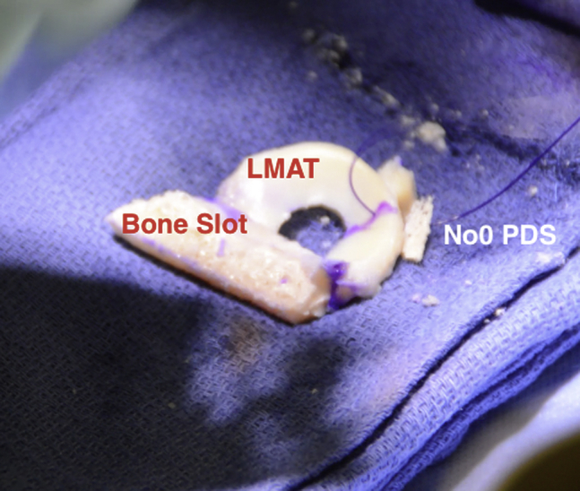
Intraoperative photograph of the lateral meniscal allograft (LMAT). The bony slot and meniscal tissue can be clearly delineated. A no. 0 PDS suture is placed through the junction of the middle and posterior third of the LMAT.
Official websites use .gov
A
.gov website belongs to an official
government organization in the United States.
Secure .gov websites use HTTPS
A lock (
) or https:// means you've safely
connected to the .gov website. Share sensitive
information only on official, secure websites.
Intraoperative photograph of the lateral meniscal allograft (LMAT). The bony slot and meniscal tissue can be clearly delineated. A no. 0 PDS suture is placed through the junction of the middle and posterior third of the LMAT.