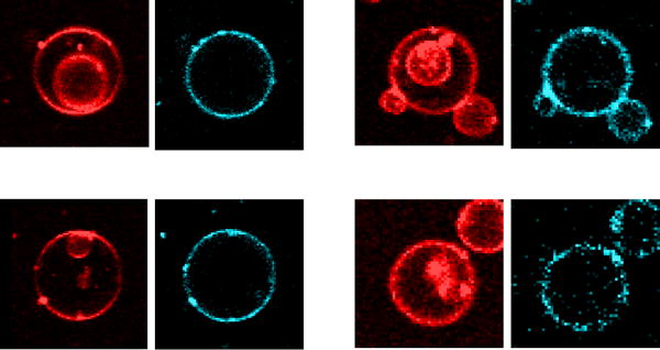
Figure 4.

Four examples of parent GUVs containing inner vesicles, in the presence of daptomycin (2 μM, added outside). No daptomycin is observed on the inner vesicle membranes. In each example, the left panel shows the fluorescence of the lipid probe LRh-DOPE (red), which was incorporated in the membrane during the GUV preparation. The right panels show the daptomycin fluorescence, from its intrinsic kynurenine chromophore (cyan). Daptomycin is present in the outer membrane of the parent GUV, but not in the inner vesicles.
