
Figure 5.
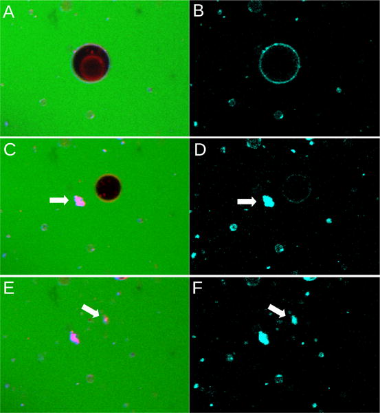
Daptomycin (2 μM) induces the collapse of a GUV composed of POPC:POPG 70:30, containing 0.1 mol% LRh-DOPE in the presence of 2 mM Ca2+. (A,B), Initial state of the same GUV. (A) All fluorescence channels (green, carboxyfluorescein; red, LRh-DOPE; cyan, daptomycin). (B) Daptomycin fluorescence channel only (cyan). (C,D) Very next image in the time scan (∼ 40 seconds between each image). The outer membrane of the GUV has now collapsed but its inner vesicle still remains (black in C). (E,F) Images taken ∼ 40 seconds later. These images were taken over a period of about 2 minutes.