
Figure 8.
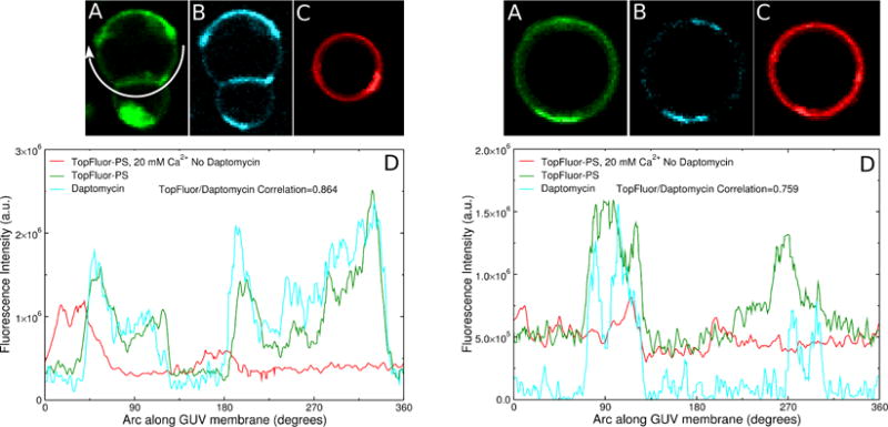
Examples of two GUVs of POPC:POPG 80:20 (left and right panels) showing TopFluor-PS and daptomycin domains in the presence of 20 mM Ca2+. The daptomycin concentration in the outside solution was 2 μM. Fluorescence of (A) TopFluor-PS and (B) daptomycin. (C) TopFluor-PS fluorescence in a GUV of POPC:POPG 80:20, without daptomycin, in the presence of 20 mM Ca2+. (D) Fluorescence intensities (same color code) along the circumference of the vesicles shown, as indicated by the arrow in (A) left panel.