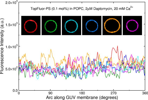
Figure 9.

Examples of six GUVs of POPC containing 1 mol% TopFluor-PS in the presence of 2 μM daptomycin and 20 mM Ca2+. The fluorescence is uniform indicating that binding of daptomycin to TopFluor-PS does not induce the domains in these experiments. Rather, POPG is required to observe domain formation. The top panel shows 6 GUVs color coded in the same manner as the fluorescence intensity lines shown in the lower panel.
