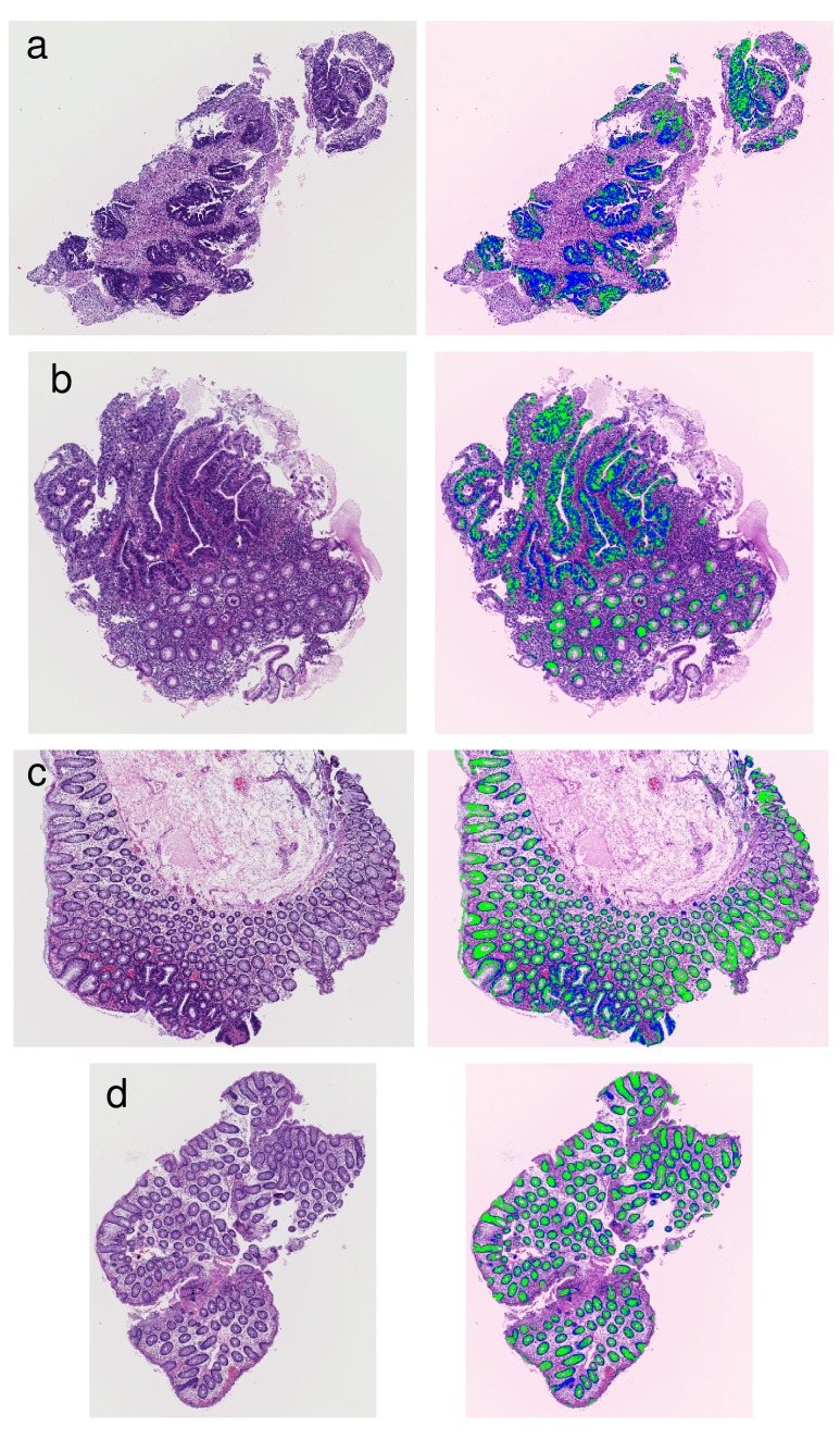
Figure 7. Examples of results that segmented glandular nuclei and glandular cytoplasm.
(a) An example of high grade atypia. (b) An example including high grade atypia. (c) An example including intermediate grade atypia. (d) An example of low grade atypia. The extracted glandular nuclei are shown in blue, and the extracted glandular cytoplasms are shown in green.

