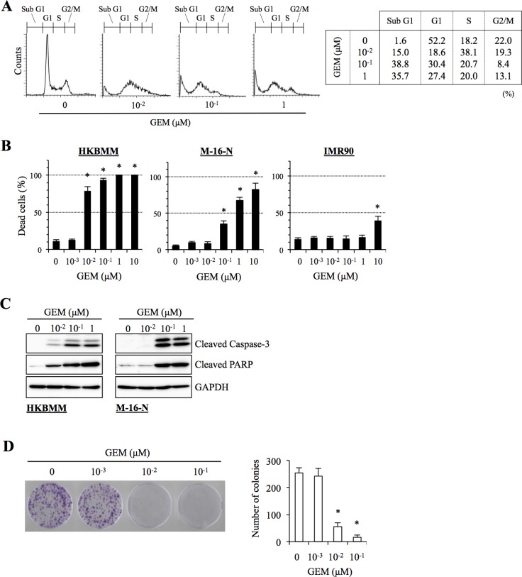
Figure 3. Anti-proliferative and pro-apoptotic activities of gemcitabine in high-grade meningioma cells.
(A) HKBMM cells treated with the indicated concentrations of gemcitabine (GEM) for 24 h were subjected to cell cycle analysis by flow cytometry. Representative flow cytometry histograms are shown, with the percentage of cells in each cell cycle phase tabulated on the right. (B) Cells treated with the indicated concentrations of GEM for 3 days were subjected to cell death assay. The graphs show the percentage of dead cells, and values in the graphs represent means + SD from triplicate samples of a representative experiment repeated with similar results. *P < 0.05. (C) Cells treated with the indicated concentrations of GEM for 24 h were subjected to immunoblot analysis of cleaved caspase-3 and PARP expression. (D) HKBMM cells treated with the indicated concentrations of GEM for 3 days were cultured for another 1 week in the absence of GEM for colony formation assay. An image of a representative experiment (left panel) and the number of colonies (right graph) are shown. Values in the graph represent means + SD from triplicate samples of a representative experiment repeated with similar results. *P < 0.05.

