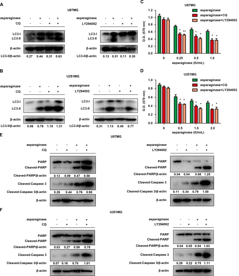
Figure 4. Abolishing autophagy potentiated asparaginase-induced growth inhibition and apoptosis of GBM cells in vitro.
(A) U87MG cells were treated with 0.5 IU/mL of asparaginase in the absence or presence of 20 μM CQ or 20 μM LY294002 for 48 h, autophagy-associated protein LC3-I/II were detected by western blot analysis. (B) U251MG cells were treated with 0.5 IU/mL of asparaginase in the absence or presence of 20 μM CQ or 10 μM LY294002 for 48 h, autophagy-associated protein LC3-I/II were detected by western blot analysis. (C) U87MG cells were treated with asparaginase at indicated concentrations in the absence or presence of 20 μM CQ or 20 μM LY294002 for 48 h, then cell viability was analyzed by MTT assay. (D) U251MG cells were treated with asparaginase at indicated concentrations in the absence or presence of 20 μM CQ or 10 μM LY294002 for 48 h, then cell viability was analyzed by MTT assay. (E) U87MG cells were treated with 0.5 IU/mL of asparaginase in the absence or presence of 20 μM CQ or 20 μM LY294002 for 48 h, then western blot analysis was performed to assess the expression level of PARP, cleaved PARP and cleaved-caspase 3. (F) U251MG cells were treated with 0.5 IU/mL of asparaginase in the absence or presence of 20 μM CQ or 10 μM LY294002 for 48 h, then western blot analysis was performed to assess the expression level of PARP, cleaved PARP and cleaved-caspase 3. Results were represented as mean ± SD (*P < 0.05, **P < 0.01).

