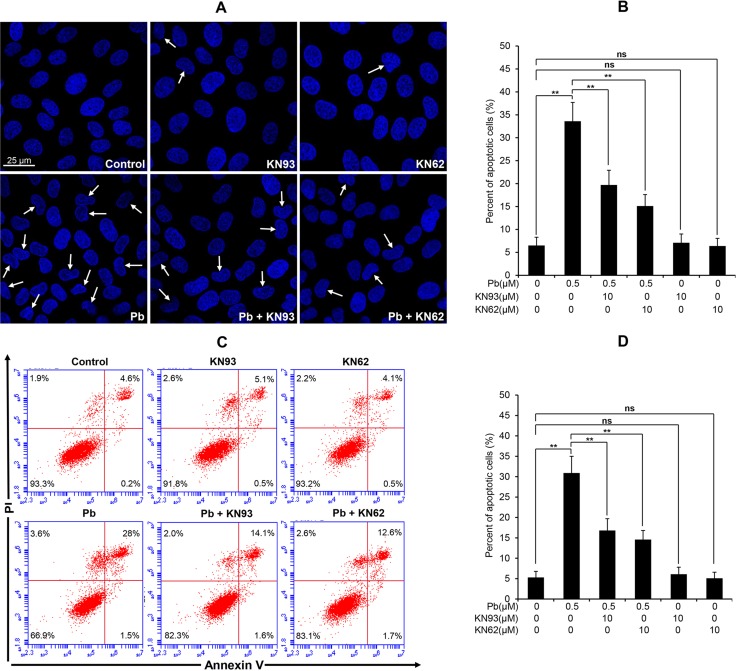
Figure 9. Effects of two CaMKII inhibitors (KN93, KN62) on Pb-induced apoptosis in rPT cells.
(A, B) Cells grown on coverslips were pre-incubated with 10 µM KN93 or 10 µM KN62 for 2 h, then exposed to 0.5 µM Pb for another 12 h to assess the apoptosis using Hoechst 33258 staining. Representative morphological changes of apoptosis are present in (A), and its statistical result of apoptotic rates (B) are expressed as mean ± SEM (n = 9). (C, D) Cells were pre-treated with 10 µM KN93 or 10 µM KN62 for 2 h, then exposed to 0.5 µM Pb for 12 h to assess the apoptosis using flow cytometry. Data in (D) are mean ± SEM of three separate experiments, and each one performed in triplicate (n = 9). ns not significant; ** P < 0.01.

