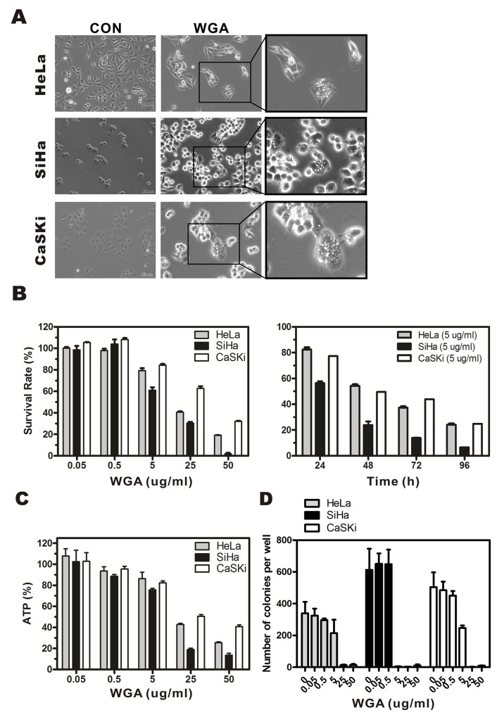
Figure 1. WGA induces formation of cytoplasmic vacuoles and paraptosis-like cell death in cervical carcinoma cells.
(A) Light micrographs of WGA-treated and untreated control cancer cells. HeLa cells, SiHa cells, and CaSKi cells were treated with WGA (10, 5, and 20 μg/mL, respectively). (B) MTT assays of cell viability. Cells were incubated with WGA at different concentrations for 24 h, or with a fixed concentration of 5 μg/mL for different lengths of time. (C) ATP levels 24 h after treatment with WGA at the indicated concentrations. (D) Clonogenic assays showing decreased viability of HeLa, SiHa, and CaSKi cells after treatment with WGA at concentrations ranging from 0.05–50 μg/mL. After long-term incubation (10–14 days), cells were fixed and stained with crystal violet, and the number of colonies counted. Data are expressed as the mean ± SD based on three independent experiments.

