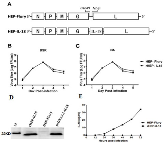
Figure 1. Construction and characterization of recombinant RABV expressing IL-18 in vitro.

(A) Schematic diagram for the construction of rHEP-IL18. The HEP-Flury vector was constructed from HEP-Flury strain by adding BsiWI and NheI sites between the G and L genes. Mouse IL-18 genes were cloned between the G and L. (B) Virus growth curves in BSR cells (B) and NA cells (C). Cells were infected with HEP-Flury or rHEP-IL18 at a multiplicity of infection (MOI) of 0.01. Viruses were harvested at 1, 2, 3, 4, 5 and 6 dpi, and viral titers were determined as described in Materials and Methods. All titrations were carried out in quadruplicate. (D) The expression of protein IL-18 was determined by Western blotting analysis. The cells were collected and lysed for Western blotting after BSR cells were infected with rHEP-IL18 or HEP-Flury for 48h. Recombinant pcDNA3.1-IL-18 was used as a positive control. (E) The expression level of murine IL18 was determined by a commercial ELISA kit. Briefly, BSR cells were infected with rHEP-IL18 or HEP-Flury (MOI=1, 0.1, 0.01, or 0.001) for 48h, and the culture supernatants were harvested for measurement of murine IL18, each value was expressed as mean ±SD from three independent experiments.
