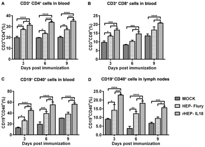
Figure 4. Recruitment and/or activation of T and B cells in blood and lymph nodes after rHEP-IL18 infection.

ICR mice were immunized i.m. with 1 × 10⁵ FFU of rHEP-IL18, HEP-Flury or DMEM. Blood and inguinal lymph nodes were collected from 3 mice per group at 3, 6, and 9 dpi. Single cell suspensions were prepared and stained with antibodies for T (CD3+, CD4+ and CD8+) from the blood (A and B) and B cells (CD19+ and CD40+) from the blood (C) and the lymph nodes (D). Asterisks indicate significant differences among the experimental groups as analyzed by one-way ANOVA (*P < 0.05;**P < 0.01;***P < 0.001;****P < 0.0001).
