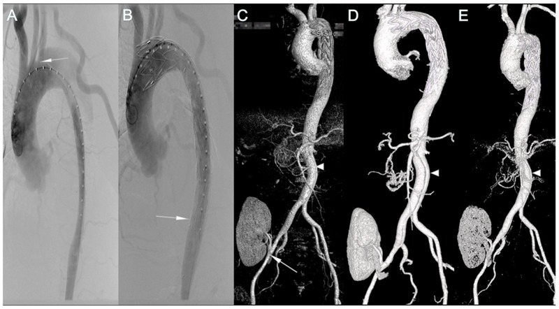
Figure 1. Pre-, Intra-, Post-operative imaging of patient 1.
(A) Aortography showed that the true lumen was compressed at the primary entry tear located at the proximal descending aorta (arrow), 5 mm to the LSA. (B) Aortography after stent deployment showed successful endovascular repair of the dissection without endoleak. The bare stent was deployed at the distal descending aorta (arrow). The lumen was expanded by the stent. (C-E) The 1st, 6th, 24th month postoperative CTA showed that none of endoleak, malperfusion of renal graft, or stenosis of renal artery was occurred (arrow), but retrograde flow from distal tear site was still existed (triangle).

