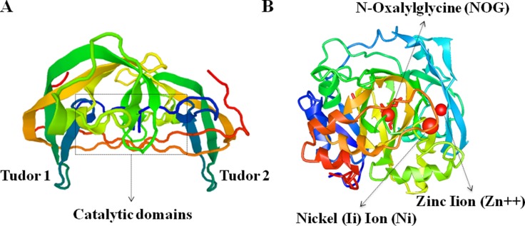
Figure 2. Crystal structure of human JMJD2C protein characterized by X-RAY diffraction.
(A) The tudor domains that recognize and bind methylated histones, and the catalytic domains are marked in the schematic image of JMJD2C crystal structures. (B) The functional moleculars including NOG (N-Oxalylglycine), Zn (Zinc Iion) and Ni (Nickel (Ii) Ion) are presented in the precise image of JMJD2C crystal structures.

