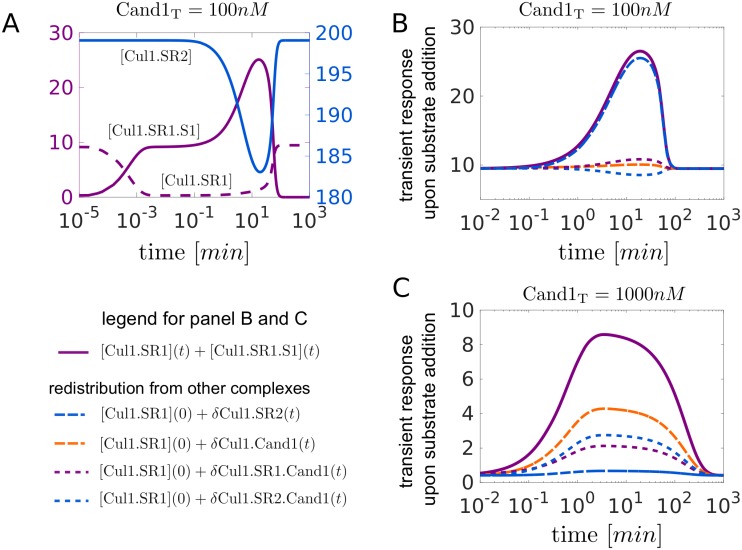
Fig 5. Dynamic readjustment of the SCF repertoire.
A: Transient response of the SCF complexes Cul1.SR1, Cul2.SR2 and Cul1.SR1.S1 upon substrate addition (300nM at t = 0) to a steady state mixture containing SR1T = 30nM, SR2T = 630nM and Cand1T = 100nM. Between 1-100min the drop in [Cul1.SR2] is accompanied by a peak in [Cul1.SR1.S1] indicating that Cul1 is redistributed from Cul1.SR2 into Cul1.SR1 and Cul1.SR1.S1. B, C: Redistribution of Cul1 from Cul1.SR2, Cul1.SR1(2).Cand1 and Cul1.Cand1 into Cul1.SR1 and Cul1.SR1.S1 for Cand1T = 100nM (B) and Cand1T = 1000nM (C). In both panels the solid violet line shows the transient increase of the concentration of “engaged” ligases ([Cul1.SR1] + [Cul1.SR1.S1]) upon substrate addition as described in (A). The remaining curves indicate the contribution to the transient response by any of the other complexes. For example, δCul1.SR2(t) = [Cul1.SR2](0) − [Cul1.SR2](t) denotes the amount of Cul1 that is redistributed into Cul1.SR1 and Cul1.SR1.S1 upon disassembly of Cul1.SR2. Note that in panels B and C the solid violet curve is the sum of the other curves. Parameters for substrate binding: kon = 108M−1s−1, koff = 1s−1, kdeg = 0.004s−1. Parameters other than those mentioned are listed in Table 1.

