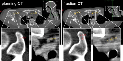
Figure 1.

Expert‐defined landmarks in a head and neck patient: (left) planning CT with two defined landmarks located in a bony structure (red cross) and next to a vascular system (yellow cross); (right) corresponding position of the same landmarks in the fraction CT independently identified by five observers. Note: To illustrate the interobserver variation in 2D, all relocated landmark positions were projected onto the same transversal slice, neglecting their real slice position.
