Figure 3.

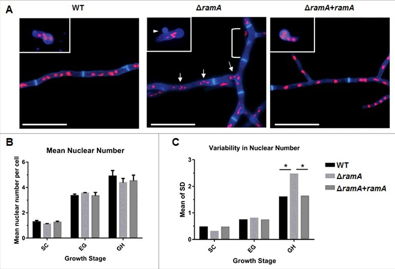
RamA is required for normal nuclear distribution in growing hyphae. (A) Nuclear positioning within germlings and hyphal compartments of the ramA isogenic set. 1 × 10⁵ conidia of each strain were inoculated onto sterile coverslips submerged in GMM broth and incubated at 37°C for various time points to enrich for selected growth stages. Incubation times were based on the germination assay results in Fig. 2 [Swollen conidia (SC): WT, ΔramA+ramA = 5.5 hours; ΔramA = 6 hours. Early germling (EG): WT, ΔramA+ramA = 6.5 hours; ΔramA = 7.5 hours. Growing hyphae (GH): all strains = 16 hours.]. Coverslips were fixed and stained with CFW to visualize the cell wall and PI to visualize nuclei. Representative micrographs of each strain are shown. Inset images are from the EG stage, whereas the main panel displays GH stage for each strain. Note the anuclear conidium, denoted by the white arrowhead, in the ΔramA EG inset micrograph. Scale bars represent 20 µm. (B) Mean nuclear number per cell of the ramA isogenic set. The number of nuclei within 20 conidia, germlings, or subapical interseptal compartments was enumerated. Nuclei were identified as discrete spots of PI staining within the cell boundaries outlined with CFW. Measurements represent the mean of 3 independent experiments ( ± SD). The mean nuclear number was not statistically different between any strains at any tested growth stage. (C) Variability of nuclear number in cells of the ramA isogenic set. The SD was calculated for each combination of strain and growth stage for each of the 3 experimental replicates in (B). Variability was defined as the overall mean of the SDs of the 3 experimental replicates for each combination of strain and growth stage. Statistics were computed by 2-way ANOVA with Tukey's test for multiple comparisons (*, p < 0.05).
