
Figure 2.
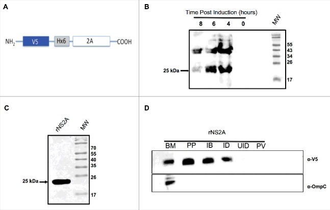
Expression and purification of NS2A protein from bacteria, and confirmation of NS2A association with bacterial membranes. (A) Schematic representation of the bacterial expression construct encoding the NS2A protein in frame with an N-terminal V5 epitope and 6 × His tag. (B) Expression and purification of NS2A protein in bacteria. Cells were induced with 1 mM IPTG at time zero, and NS2A expression was analyzed by western blot at the indicated times post-induction using an anti-V5 antibody (dilution, 1:5000). (C) Analysis of recombinant NS2A protein (rNS2A) purification following different isolation steps by SDS-PAGE and Coomassie blue staining. Preparative gel electrophoresis was used for purification. Lane 1: purified fraction of NS2A obtained from preparative gels (molecular-weight, approximately 25 kDa). (D) Localization of NS2A within Escherichia coli membranes was detected via western blot analysis using an anti-V5 antibody and a mouse polyclonal antibody specific for OmpC (a bacterial membrane protein). Lane 1: purified bacterial membranes (BM); lane 2: purified NS2A protein (PP); lane 3: inclusion bodies of bacteria transformed with NS2A (IB); lane 4: NS2A clone, induced with IPTG (ID); lane 5: NS2A clone, uninduced (UID); lane 6: bacteria transformed with the parental vector (PV).