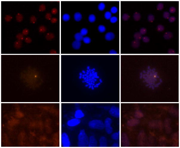
Figure 13.
Detection of chromosomal DNA using Invader probes. Fluorescence micrographs from fluorescence in situ hybridization experiments using Y-chromosome specific Invader probes under non-denaturing conditions. Invader probe 5′-Cy3-AGCCCUGTGCCCTG:3′-TCGGGACACGGGAC-Cy3 was incubated with nuclei from male bovine kidney cells in interphase (upper panel) or metaphase (middle panel), or with nuclei from female bovine fibroblast cells (lower panel). Images viewed using Cy3 (left column) or DAPI (middle column) filter settings; overlays are shown in the right column. Adapted from reference 54 with permission from the Royal Society of Chemistry.

