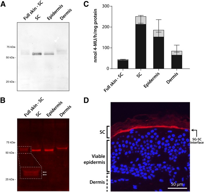
Fig. 2.
Labeling of GBA1 in human skin tissue. A: Western blot of GBA1 in different human skin tissue layers using monoclonal anti-GBA1 (50 μg of protein per lane were loaded on the SDS-PAGE gel). Full skin-SC, full thickness skin without SC. B: Fluorescent labeling of active GBA1 (5 μg of protein per lane were loaded on the SDS-PAGE gel) in skin tissues exposed to MDW941. C: Bar plots of the enzymatic activity of GBA in skin tissue, as determined by a 4-MU-β-glc substrate assay (gray + black bars). The fraction of 4-methylumbelliferone converted by GBA1 is indicated by black bars (n = 3, mean ± SD). D: Immunohistochemical fluorescent staining of expressed GBA1 (red), and counterstaining with DAPI (blue) for cell nuclei. Objective lens magnification 20×.

