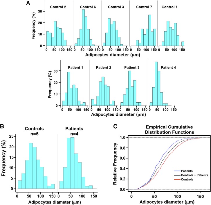
Fig. 3.
Histomorphometric analysis of adipocyte diameter in patients carrying biallelic LPIN1 inactivating mutations. The diameter (expressed in μm) of approximately 100 adipocytes was measured in each subcutaneous adipose tissue histological section from five normal controls and four lipin-1-deficient patients. Control 1, age 12; Control 2, age 15; Control 3, age 18; Control 6, age 15; Control 7, age 15; Patient 1, age 5; Patient 2, age 5; Patient 3, age 11; Patient 4, age 47 (see Table 1). The controls were highly homogeneous in age (controls: n = 5, mean age: 15; SD = 1.9), and treated as a group. Two different statistical approaches were used to minimize the possible confounding effect of age on adipocyte size (see Results section and Tables 2, 3). A: histograms of the frequency distribution of adipocyte diameter for each control and each patient. B: histogram of the frequency distribution of adipocyte diameter for the control group (n = 5) and the patient group (n = 4). C: ECDFs of adipocytes diameter for all controls (red), all patients (blue), and the total sample (controls + patients) (black). These experiments (see Results section and Tables 2, 3) show a significant decrease in adipocyte size in patients compared with normal controls.

