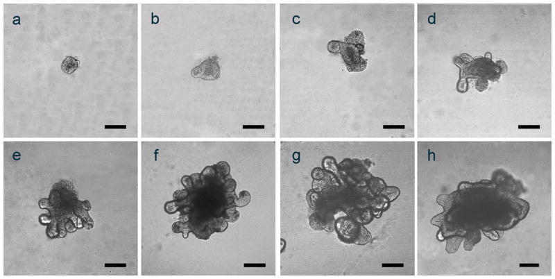
Figure 1. Representative images of small intestinal (jejunum) and colonic organoids.
Confocal microscopic images were taken of representative small intestinal (days 0–7; a–g) and colonic organoids (day 21, h) growing in 24 well plates as described in Methods. Scale bars in all panels (a–h) represent 100μm.

