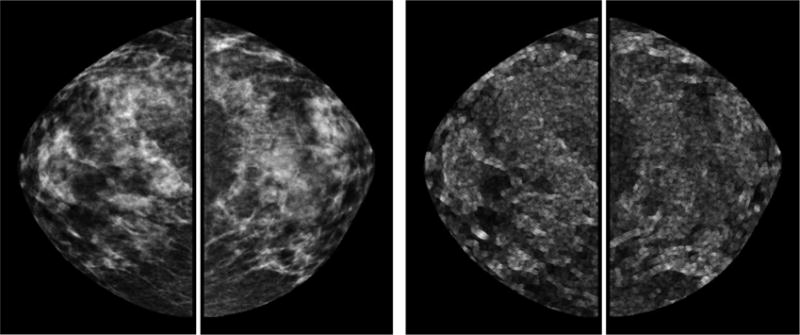
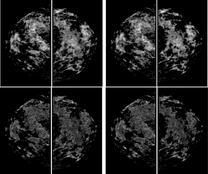
Fig. 1.


Example of a positive case acquired from the “prior” screening examination. It shows the segmented whole breast regions of original images (top left) and local pixel value fluctuation maps (top right), dense breast regions of original images segmented with the new segmentation method (middle left) and previous segmentation method (middle right), dense breast regions of local pixel value fluctuation maps segmented with the new segmentation method (bottom left) and previous segmentation method (bottom right).
