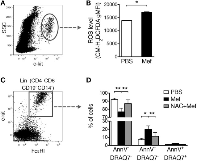
Figure 4.

Oxidative stress induced by mefloquine is a major cause of mast cell apoptosis. (A,B) Mixed human lung cells were prepared by mechanical and enzymatic treatment of lung specimens and treated with phosphate-buffered saline (PBS) or mefloquine (20 µM) for 1 h. Next, c-kit+ cells were gated (A) and the reactive oxygen species (ROS) levels were quantified using the fluorescent ROS probe CM-H2DCFDA (B). The bars in panel (B) represent mean ± SEM of the geometric mean fluorescence intensity (gMFI) for the CM-H2DCFDA (*P < 0.05) (n = 2; representative of two independent experiments/two donors). (C,D) Mixed human lung cells were pre-incubated with or without N-acetylcysteine (NAC) (8 mmol/L) for 2 h, followed by treatment with mefloquine (20 µM) or PBS for 24 h. Lin− c-kit+ FcεRI+ mast cells were gated (C) and the percentage of viable (Annexin V−/DRAQ7−), apoptotic (Annexin V+/DRAQ7−) and necrotic (Annexin V+/DRAQ7+) mast cells were determined (D) (n = 2; representative of three independent experiments/three donors). Data are presented as mean ± SEM (*P < 0.05, **P < 0.01).
