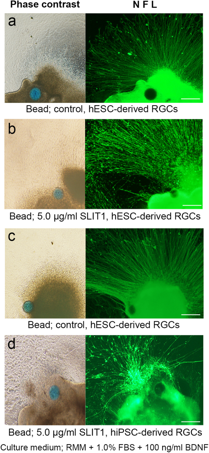
Figure 8.

The effect of focally sustained release of SLIT1 on pathfinding of axons derived from hESC- and hiPSC-derived RGCs. Phase-contrast images showing SLIT1-releasing beads (blue-coloured) placed next to the bottom of attached OV, and corresponding immunohistochemistry for the axons of the RGCs by NFL staining. (a,c) The paths of the axons of hESC- and hiPSCs- derived RGCs ((a) and (c), respectively) stained by NFL radiate straight from the attached OV, even though the bead is located close by. (b,d) Compared with the control, the axons of hESC- and hiPSC-derived RGCs ((b) and (d), respectively) are repelled by SLIT1 release from the bead, and axonal paths are not straight, but winding, and avoid the beads. The assessment from D27 is performed in RMM supplemented with 1.0% FBS and 100 ng/ml BDNF. Each experiment was repeated at least five times. Scale bars, 100 μm.
